Comparison of Unmonochromatized Synchrotron Radiation and Conventional X-rays in the Imaging of Mammographic Phantom and Human Breast Specimens: A Preliminary Result
- Affiliations
-
- 1Department of Diagnostic Radiology, Yonsei Univercity College of Medicine, Seoul, Korea. hjkim@yumc.yonsei.ac.kr
- 2Research Institute of Radiological Science, Yonsei Univercity College of Medicine, Seoul, Korea.
- 3BK21 Project for Medical Sciences, Yonsei Univercity College of Medicine, Seoul, Korea.
- 4Department of Materials Science and Engineering, Pohang Univercity of Sci. and Technol., Pohang, Korea.
- 5Institute of Physics, Academia Sinica, Nankang 11529, Taipei, Taiwan.
- 6Institut de physique Appliquee, Ecole Polytechnique Federale de Lausanne, CH-1015 Lausanne, Switzerland.
- KMID: 2158118
- DOI: http://doi.org/10.3349/ymj.2005.46.1.95
Abstract
- A simple imaging setup based on the principle of coherence-based contrast X-ray imaging with unmonochromatized synchrotron radiation was used for studying mammographic phantom and human breast specimens. The use of unmonochromatized synchrotron radiation simplifies the instrumentation, decreases the cost and makes the procedure simpler and potentially more suitable for clinical applications. The imaging systems consisted of changeable silicon wafer attenuators, a tungsten slit system, a CdWO4 scintillator screen, a CCD (Charge Coupled Device) camera coupled to optical magnification lenses, and a personal computer. In preliminary studies, a spatial resolution test pattern and glass capillary filled with air bubbles were imaged to evaluate the resOolution characteristics and coherence-based contrast enhancement. Both the spatial resolution and image quality of the proposed system were compared with those of a conventional mammography system in order to establish the characteristic advantages of this approach. The images obtained with the proposed system showed a resolution of at least 25micrometer on the test pattern with much better contrast, while the images of the capillary filled with air bubbles revealed coherence-based edge enhancement. This result shows that the coherence-based contrast imaging system, which emphasizes the refraction effect from the edge of materials of different refractive indexes, is applicable to imaging studies in fundamental medicine and biology, although further research works will be required before it can be used for clinical applications.
Keyword
MeSH Terms
Figure
-
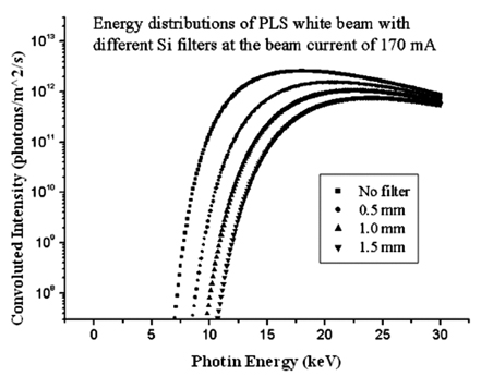
Fig. 1 Energy spectra of PLS white beam for different silicon wafers thicknesses. A series of attenuators was placed in the beam upstream of the sample position and the energy range and the photon flux were adjusted in the range of 6-25 keV and 1011 - 1012 photons/mm2/sec, respectively.
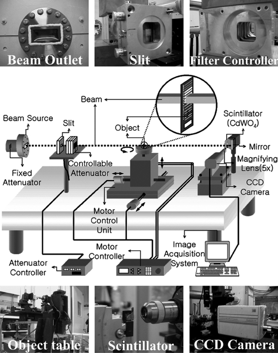
Fig. 2 Schematic diagram of unmonochromatized synchrotron radiation imaging set-up at PLS 5C1 beamline. Synchrotron radiation X-rays passing through the test objects then interact with the scintillator screen before being converted to a visible light image. The optical images were magnified by optical lenses, captured by a CCD camera and transferred to a computer for data acquisition and analysis.
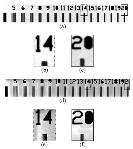
Fig. 3 The images of the spatial resolution test pattern; (a) complete image, (b) small input field of 14 line pairs mm-1, and (C) small input field of 20 line pairs mm-1, obtained by the conventional mammography system (GE Senographe DMR) and (d) complete image, (e) small input field of 14 line pairs mm-1, and (f) small input field of 20 line pairs mm-1, obtained by the PLS 5C1 synchrotron imaging system. The PLS 5C1 unmonochromatized synchrotron imaging system provided a resolution of at least 20 line-pairs mm-1 (25 µm) with the resolution test pattern compared to the 14 line-pairs mm-1 (≈36 µm) spatial resolution obtained with the conventional mammography system.
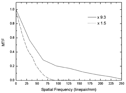
Fig. 4 The modulation transfer function (MTF) of the PLS 5C1 SR imaging system at 1.5 × and 9.5 × optical magnifications for the horizontal direction in relation to the incident beam. The spatial resolution (10% of MTF) was found to be about 60 line pairs/mm (corresponds to 8.3 nm) for the imaging setup with 1.5 × optical magnification.
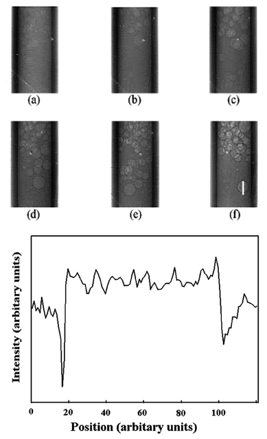
Fig. 5 The PLS 5C1 unmonochromatized synchrotron radiation images of a glass capillary tube filled with air bubbles for various object-to-detector (scintillator) distances viz. for (a) to (f), 50, 100, 200, 250, 300, and 470 mm, respectively. The sequential images illustrate the effect of the coherence-based contrast which operates by the mechanism of refraction-based edge enhancement within a certain range of object-detector distance values. (g) Cross-sectional intensity profile of the bubble along the vertical line in (f) (arbitrary units scaled to each other).

Fig. 6 Schematic and images of the mammographic accreditation phantom (Gammex RMI 156); (a) a schematic view of the mammographic phantom giving the test object sizes and positions, (b) radiographic image obtained with the conventional mammographic system, (c) partial image of artificial microcalcification obtained with the conventional mammography system, (d) partial image of artificial microcalcification obtained with the PLS 5C1 synchrotron imaging system. The synchrotron image of artificial microcalcification shows more details and clearer boundaries.
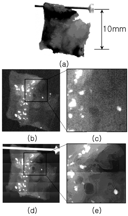
Fig. 7 Images of human breast cancer specimens preserved in formalin acquired from conventional mammographic system and the PLS 5C1 unmonochromatized synchrotron imaging system; (a) gross photographic image of specimen, (b) whole image, and (c) small input field of image b obtained with the conventional mammography system, (d) complete image, and (e) small input field of image obtained with the synchrotron radiation imaging system. In the images (b)-(e), white spots show the images of the microcalcifications detected in the breast cancer tissue. In the synchrotron radiation images, the contrast was enhanced and the details were depicted more sharply than in the conventional images. In particular, the edges of the calcification and mass were well defined. Here, the synchrotron radiation images were inverted for the purpose of comparison with the mammographic images. (In the synchrotron radiation images, the upper and right white bars indicate the needle used for hanging the specimen during image acquisition.)
Cited by 1 articles
-
A New Method for Investigation of the Hair Shaft: Hard X-Ray Microscopy with a 90-nm Spatial Resolution
Soo-Young Jeon, Ja Woong Goo, Seung Phil Hong, Tak Heon Oh, Hwa Shik Youn, Won-Soo Lee
Yonsei Med J. 2008;49(2):337-340. doi: 10.3349/ymj.2008.49.2.337.
Reference
-
1. Momose A, Fukuda J. Phase-contrast radiographs of nonstained rat cerebellar specimen. Med Phys. 1995. 22:375–379.2. Davis TJ, Gao D, Gureyev TE, Stevenson AW, Wilkins SW. Phase-contrast imaging of weakly absorbing materials using hard x-rays. Nature. 1995. 373:595–598.3. Snigirev A, Snigireva I, Kohn V, Kuznetsov S, Schelokov I. On the possibilities of x-ray phase contrast microimaging by coherent high-energy synchrotron radiation. Rev Sci Instrum. 1995. 66:5486–5492.4. Wilkins SW, Gureyev TE, Gao D, Pogany A, Stevension AW. Phase-contrast imaging using polychromatic hard x-rays. Nature. 1996. 384:335–338.5. Chapman D, Thomlinson W, Johnston RE, Washburn D, Pisano ED, Gmur NF, et al. Diffraction enhanced x-ray imaging. Phys Med Biol. 1997. 42:2015–2025.6. Gao D, Pogany A, Stevenson AW, Wilkins SW. Phase-contrast radiography. Radiographics. 1998. 18:1257–1267.7. Margaritondo G, Tromba G. Coherence-based edge diffraction sharpening of x-ray images: a simple model. J Appl Phys. 1999. 85:3406–3408.8. Hwu Y, Hsieh HH, Lu MJ, Tsai WL, Lin HM, Goh WC, et al. Coherence-enhanced synchrotron radiation: refraction versus diffraction mechanisms. J Appl Phys. 1999. 86:4613–4618.9. Kim HJ, Hong JO, Lee KH, Jung H, Kim EK, Je JH, et al. Phantom and animal imaging studies using PLS synchrotron x-rays. IEEE T Nucl Sci. 2001. 48:837–842.10. Hwu Y, Je JH, Lee KH, Margaritondo G. Real time micro-radiology with SR on live specimens. In : 7th International Conference on Synchrotron Radiation Instrumentation; Book of Abstracts, THU2-01 invited 2000.11. Hwu Y, Tsai WL, Hsieh HH, Je JH, Kang HS, Kim IW, et al. Collimation-enhanced micro-radiography in realtime. Nucl Instrum Meth A. 2001. 467-8:294–1300.12. Yoneyama A, Momose A, Seya E, Hirano K, Takeda T, Itai Y. Operation of a separated-type x-ray interferometer for phase-contrast x-ray imaging. Rev Sci Instrum. 1999. 70:4582–4586.13. Takeda T, Momose A, Hirano K, Haraoka S, Watanabe T, Itai Y. Human carcinoma: early experience with phase-contrast x-ray CT with synchrotron radiation-comparative specimen study with optical microscopy. Radiology. 2000. 214:298–301.14. Pisano ED, Johnston RE, Chapman D, Geradts J, Lacocca MV, Livasy CA, et al. Human breast cancer specimens: diffraction-enhanced imaging with histologic correlation-improved conspicuity of lesion detail compared with digital radiography. Radiology. 2000. 214:895–901.15. Jung H, Kim HJ, Hong S, Hong JO, Jeong HK, Je JH, et al. Computed microtomography (µCT) with unmonochromatized synchrotron X-rays for cancerous human breast tissue and mouse vertebra. IEEE T Nucl Sci. 2002. 49:2262–2267.16. Hwu Y, Lai B, Mancini DC, Je JH, Noh DY, Bertolo M, et al. Coherence based contrast enhancement in x-ray radiography with a photoelectric microscope. Appl Phys Lett. 1999. 75:2377–2379.17. Umetani K, Yagi N, Suzuki Y, Kohmura Y, Yamasaki K. X-ray refraction-contrast imaging using synchrotron radiation at Spring-8. Proceedings of SPIE. 1999. 3659:In : Medical Imaging 1999: Physics of Medical Imaging; 560–571.18. Yu Q, Takeda T, Umetani K, Ueno E, Itai Y, Hiranaka Y, et al. First experiment by two-dimensional digital mammography with synchrotron radiation. J Synchrotron Rad. 1999. 6:1148–1152.19. Arfelli F, Bonvicini V, Bravin A, Cantatore G, Castelli E, Palma LD, et al. Mammography with synchrotron radiation: phase-detection techniques. Radiology. 2000. 215:286–293.20. Burattini E, Cossu E, Maggio CD, Gambaccini M, Indovina PL, Marziani M, et al. Mammography with synchrotron radiation. Radiology. 1995. 195:239–244.
- Full Text Links
-
- Actions
-
Cited
- CITED
-
- Close
- Share
-
- Similar articles
-
- Synchrotron Radiation Imaging of Internal Structures in Live Animals
- Synchrotron Radiation Imaging of Breast Tissue Using a Phase-contrast Hard X-ray Microscope
- Synchrotron Radiation Imaging of Female Breast Tissues Using Phase Contrast Technique
- Comparison of Image Quality between Mammography Dedicated Monitor and UHD 4K Monitor, Using Standard Mammographic Phantom: A Preliminary Study
- Usefulness of a Small-Field Digital Mammographic Imaging System Using Parabolic Polycapillary Optics as a Diagnostic Imaging Tool: a Preliminary Study
