Primary Malignant Rhabdoid Melanoma
- Affiliations
-
- 1Department of Dermatology, College of Medicine, Hallym University, Seoul, Korea. dermlee@yahoo.co.kr
- KMID: 2156778
- DOI: http://doi.org/10.5021/ad.2011.23.S2.S155
Abstract
- Melanoma with rhabdoid features is an uncommon variant of malignant melanoma. Here, we describe a rare case of primary rhabdoid malignant melanoma. A 54-year-old man presented with a black tumor measuring 3x4 cm on the right forearm. Histologic sections showed a tumor mass with rhabdoid features composed entirely of polygonal neoplastic cells with eccentric nuclei, prominent nucleoli, and large hyaline cytoplasmic inclusions. The tumor cells were immunoreactive with HMB-45, S100, Fontana-Masson silver and vimentin, and negative for smooth muscle actin, CD68, CD34, CD99, synaptophysin, desmin, and PAS. The differential diagnosis for this tumor included malignant peripheral nerve sheath tumor, malignant peripheral neuroectodermal tumor and rhabdomyosarcoma. The patient was treated with a wide excision and a local skin graft. The excised tumor was entirely composed of rhabdoid tumor cells. No recurrence or metastasis was evident 4 months after removal. This article is relevant to rare cases of primary malignant melanomas showing rhabdoid tumor cells over the entire excised lesion.
Keyword
MeSH Terms
-
Actins
Desmin
Diagnosis, Differential
Forearm
Humans
Hyalin
Inclusion Bodies
Melanoma
Middle Aged
Muscle, Smooth
Neoplasm Metastasis
Neuroectodermal Tumors, Primitive, Peripheral
Peripheral Nerves
Recurrence
Rhabdoid Tumor
Rhabdomyosarcoma
Silver
Skin
Synaptophysin
Transplants
Vimentin
Actins
Desmin
Silver
Synaptophysin
Vimentin
Figure
-
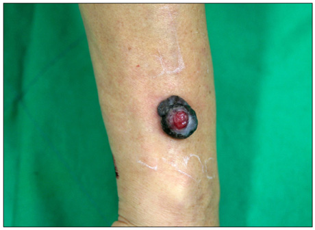
Fig. 1 A solitary, well-defined sessile black tumor measuring 3×4 cm with a granulating protrusion in the central area of the right forearm.
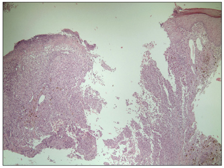
Fig. 2 An ulcerated tumor nodule showed neoplastic proliferation of polygonal cells with rhabdoid features and scattered melanin pigments (H&E, ×40).
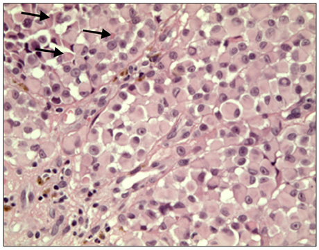
Fig. 3 Tumor cells exhibited eosinophilic cytoplasm displacing a round eccentric vesicular nucleus with a prominent central nucleolus and a large intracytoplasmic hyalin inclusion (arrows) (H&E, ×400).
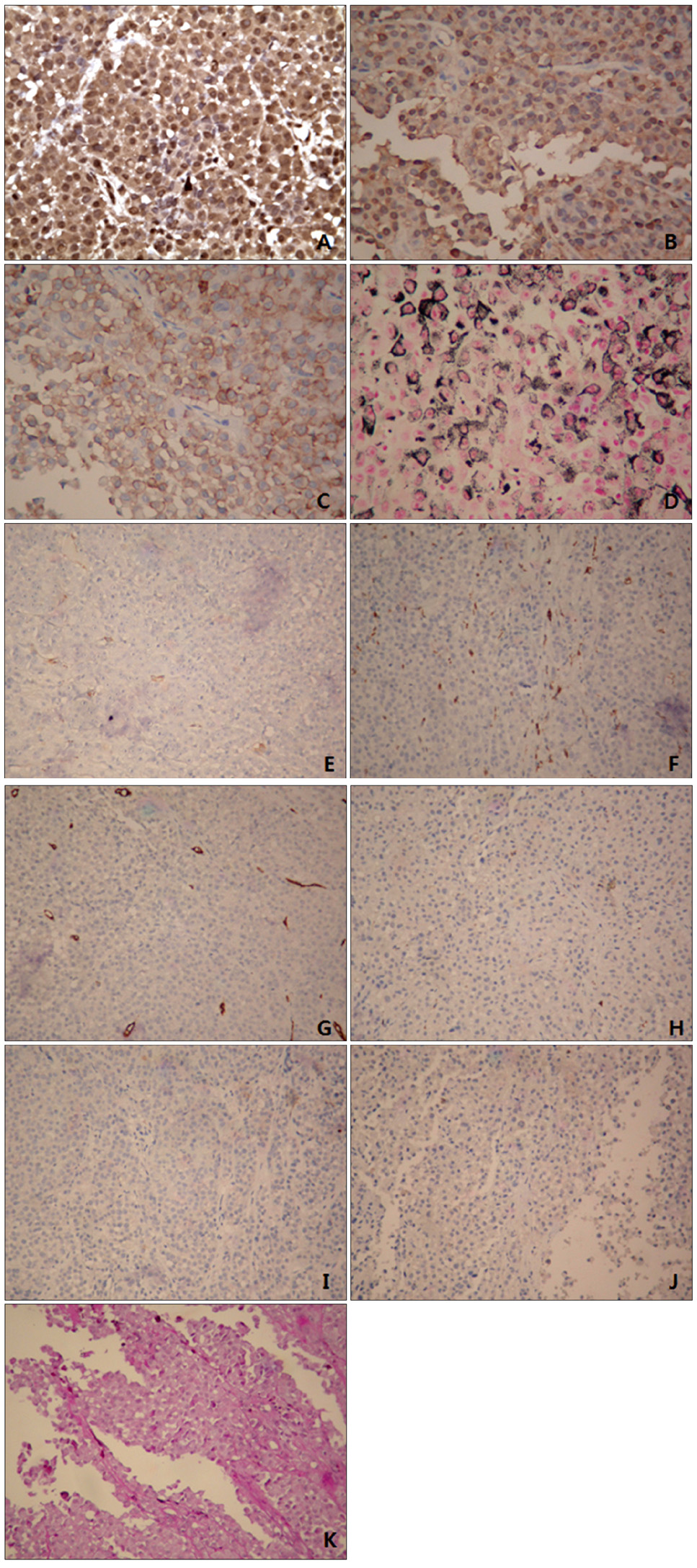
Fig. 4 Immunohistochemically, the neoplastic cells were positive for vimentin (A), S100 (B), Fontana-Masson silver (C) and HMB-45 (D), and negative for smooth muscle actin (E), CD68 (F), CD34 (G), CD99 (H), synaptophysin (I), desmin (J), and PAS (K). Vimentin staining showed intense, diffuse, and globular perinuclear accentuations (A) (A~D: H&E, ×400, E~K: H&E, ×200).
Reference
-
1. Haas JE, Palmer NF, Weinberg AG, Beckwith JB. Ultrastructure of malignant rhabdoid tumor of the kidney. A distinctive renal tumor of children. Hum Pathol. 1981. 12:646–657.2. Schmidt D, Harms D, Zieger G. Malignant rhabdoid tumor of the kidney. Histopathology, ultrastructure and comments on differential diagnosis. Virchows Arch A Pathol Anat Histopathol. 1982. 398:101–108.
Article3. Gavino AC, Gillies EM. Metastatic rhabdoid melanoma: report of a case with a comparative review of the literature. J Cutan Pathol. 2008. 35:337–342.
Article4. Bittesini L, Dei Tos AP, Fletcher CD. Metastatic malignant melanoma showing a rhabdoid phenotype: further evidence of a non-specific histological pattern. Histopathology. 1992. 20:167–170.
Article5. Magro CM, Crowson AN, Mihm MC. Unusual variants of malignant melanoma. Mod Pathol. 2006. 19:Suppl 2. S41–S70.
Article6. Chang ES, Wick MR, Swanson PE, Dehner LP. Metastatic malignant melanoma with "rhabdoid" features. Am J Clin Pathol. 1994. 102:426–431.
Article7. Parham DM, Weeks DA, Beckwith JB. The clinicopathologic spectrum of putative extrarenal rhabdoid tumors. An analysis of 42 cases studied with immunohistochemistry or electron microscopy. Am J Surg Pathol. 1994. 18:1010–1029.
Article8. Borek BT, McKee PH, Freeman JA, Maguire B, Brander WL, Calonje E. Primary malignant melanoma with rhabdoid features: a histologic and immunocytochemical study of three cases. Am J Dermatopathol. 1998. 20:123–127.
Article9. Laskin WB, Weiss SW, Bratthauer GL. Epithelioid variant of malignant peripheral nerve sheath tumor (malignant epithelioid schwannoma). Am J Surg Pathol. 1991. 15:1136–1145.
Article10. Weeks DA, Beckwith JB, Mierau GW. Rhabdoid tumor. An entity or a phenotype? Arch Pathol Lab Med. 1989. 113:113–114.11. Gattenlöhner S, Brocker EB, Muller-Hermelink HK. Malignant melanoma with metastatic rhabdomyosarcomatoid transdifferentiation. N Engl J Med. 2008. 358:649–650.
Article12. Tallon B, Bhawan J. Primary rhabdoid melanoma with clonal recurrence. Am J Dermatopathol. 2009. 31:200–204.
Article13. Abbott JJ, Amirkhan RH, Hoang MP. Malignant melanoma with a rhabdoid phenotype: histologic, immunohistochemical, and ultrastructural study of a case and review of the literature. Arch Pathol Lab Med. 2004. 128:686–688.
Article
- Full Text Links
-
- Actions
-
Cited
- CITED
-
- Close
- Share
-
- Similar articles
-
- Primary malignant melanoma arising in a cystic teratoma
- A Case of Primary Malignant Melanoma of the Vagina: Trial of a Wide Local Excision of Vagina and Rectum
- Imaging Findings of a Malignant Rhabdoid Tumor in the Stomach: A Case Report
- Primary Malignant Melanoma of the Cervical Spinal Nerve Root: A Case Report
- Primary malignant melanoma of the esophagus
