Evaluation of Engraftment of Superparamagnetic Iron Oxide-Labeled Mesenchymal Stem Cells Using Three-Dimensional Reconstruction of Magnetic Resonance Imaging in Photothrombotic Cerebral Infarction Models of Rats
- Affiliations
-
- 1Department of Convergence Medicine and Pharmaceutical Biosciences, Chung-Ang University, Seoul 156-756, Korea.
- 2Department of Radiology, Chung-Ang University Hospital, Chung-Ang University College of Medicine, Seoul 156-755, Korea. kwakbk@cau.ac.kr
- 3Major of Biomedical Science, Chung-Ang University College of Medicine, Seoul 156-756, Korea.
- KMID: 2155527
- DOI: http://doi.org/10.3348/kjr.2015.16.3.575
Abstract
OBJECTIVE
To evaluate engraftment by visualizing the location of human bone marrow-derived mesenchymal stem cells (hBM-MSCs) three-dimensionally in photothrombotic cerebral infarction (PTCI) models of rats.
MATERIALS AND METHODS
Magnetic resonance imaging (MRI) of an agarose block containing superparamagnetic iron oxide (SPIO)-labeled hBM-MSCs was performed using a 3.0-T MRI, T2-(T2WI), T2*-(T2*WI), and susceptibility-weighted images (SWI). PTCI was induced in 6 rats, and 2.5 x 10(5) SPIO-labeled hBM-MSCs were infused through the ipsilateral internal carotid artery (ICA group) or tail vein (IV group). MRI was performed on days 1, 3, 7, and 14 after stem cell injection. Dark signal regions were confirmed using histology. Three-dimensional MRI reconstruction was performed using the clinical workflow solution to evaluate the engraftment of hBM-MSCs. Volumetric analysis of the engraftment was also performed.
RESULTS
The volumes of SPIO-labeled hBM-MSCs in the phantom MRI were 129.3, 68.4, and 25.9 microL using SWI, T2*WI, and T2WI, respectively. SPIO-labeled hBM-MSCs appeared on day 1 after injection, encircling the cerebral infarction from the ventral side. Dark signal regions matched iron positive cells and human origin (positive) cells. The volume of the engraftment was larger in the ICA group on days 1, 3, and 7, after stem cell injection (p < 0.05 on SWI). SWI was the most sensitive MRI pulse sequence (p < 0.05). The volume of infarction decreased until day 14.
CONCLUSION
The engraftment of SPIO-labeled hBM-MSCs can be visualized and evaluated three-dimensionally in PTCI models of rats. The engraftment volume was larger in the ICA group than IV group on early stage within one week.
Keyword
MeSH Terms
-
Animals
Cerebral Infarction/pathology/*radiography
Contrast Media
Dextrans
Humans
Imaging, Three-Dimensional/methods
Magnetic Resonance Imaging/*methods
Magnetite Nanoparticles
Male
*Mesenchymal Stem Cell Transplantation
Mesenchymal Stromal Cells/radiography
Nanoparticles
Neuroimaging/*methods
Random Allocation
Rats
Rats, Sprague-Dawley
Tomography, X-Ray Computed
Contrast Media
Dextrans
Magnetite Nanoparticles
Figure
-
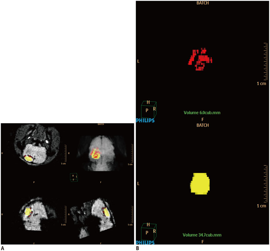
Fig. 1 Three-dimensional reconstruction of MR images of photothrombotic cerebral infarction and superparamagnetic iron oxide (SPIO)-labeled stem cells in rat model. A. Using Philips clinical workflow solution, infarction area was assigned color yellow, and dark region due to SPIO-labeled human bone marrow-derived mesenchymal stem cells was assigned color red. Three-dimensional reconstruction was performed using volume-rendering mode. B. Subsequently, volumes of infarction and SPIO were obtained (mm3) using calculate-volume tool.

Fig. 2 Confirmation of labeling stem cells with superparamagnetic iron oxide (SPIO) nanoparticles. Un-labeled human bone marrow-derived mesenchymal stem cells (hBM-MSCs) (A) and SPIO-labeled hBM-MSCs at 50 µg Fe/mL (B) stained with Perls' reagent. All cells in B were sufficiently labeled without any abnormal changes in morphology (× 400, Prussian blue staining).

Fig. 3 Two-dimensional (2D) and three-dimensional (3D) reconstruction of phantom MR images. Solidified agarose phantom had 2.2-mm diameter U-shaped column with 6.0 × 10² superparamagnetic iron oxide (SPIO)-labeled stem cells in total volume of 200-µL. A. Column is seen as U-shaped dark signal region on T2-weighted images (T2WI), T2*-weighted images (T2*WI), and susceptibility-weighted images (SWI). Dark signal region is large and prominent on SWI, followed by T2*WI, and then T2WI. B. In three-dimensionally reconstructed images, color red was assigned to dark signal region due to SPIO-labeled human bone marrow-derived mesenchymal stem cells. Volumes were 25.9, 68.4, and 129.3 µL on T2WI, T2*WI, and SWI, respectively.

Fig. 4 Time-elapsed MR images of photothrombotic cerebral infarction and superparamagnetic iron oxide (SPIO)-labeled stem cells in rat model. MR images were obtained on day 2 after photothrombotic cerebral infarction (pre, 1 day before cell injection) and on days 1, 3, 7, and 14 after internal carotid arterial injection of SPIO-labeled human bone marrow-derived mesenchymal stem cells. As time progressed, high signal intensity of infarction (⊚) was reduced in size and intensity, and dark signal region (arrowheads) appeared by encircling infarction from day 1 until day 14, indicating engraftment of stem cells to infarction. Dark regions were large and prominent in susceptibility-weighted images (SWI), followed by T2*-weighted images (T2*WI), and then by T2-weighted images (T2WI).

Fig. 5 Correlation between MR image and histology. Dark signal region (A) encircling lateral margin of cerebral infarction (⊚) on MR image (day 14) was correlated with hematoxylin and eosin (HE) staining (B, × 12.5), Prussian blue (PB) staining (C, × 100), and immunohistochemistry (IHC) with anti-human mitochondria antibody (D, × 100). On HE staining, infarction (⊚) and cystic encephalomalacia (**) were seen in right parietal lobe (A, B). On PB staining and IHC, dark regions on MR image were confirmed as superparamagnetic iron oxide-labeled human bone marrow-derived mesenchymal stem cells (A, C, D).
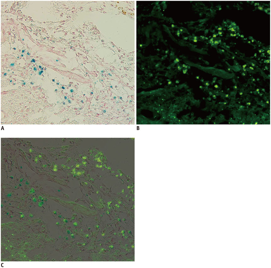
Fig. 6 Correlation between Prussian blue and immunohistochemistry (IHC) staining. Two images of Prussian blue staining (A, × 200) and IHC with anti-human mitochondria antibody (B, × 200) of same slide are superimposed (C). Positive cells can be seen at periphery of cerebral infarction. Blue-color stained iron positive cells (A) were thoroughly matched to green fluorescent human mitochondria positive cells (B). Engraftment of implanted superparamagnetic iron oxide-labeled human bone marrow-derived mesenchymal stem cells was confirmed by matching.
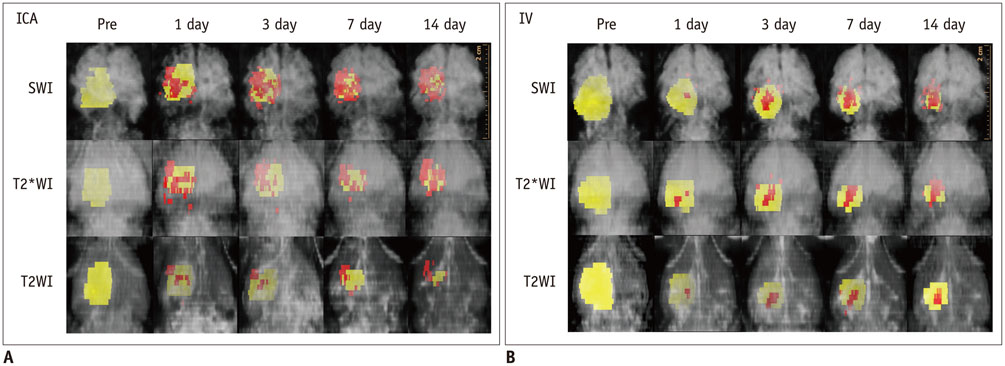
Fig. 7 Ventral perspective of three-dimensional reconstruction of MR images of photothrombotic cerebral infarction and superparamagnetic iron oxide (SPIO)-labeled human bone marrow-derived mesenchymal stem cells (hBM-MSCs) in rats. Color yellow was assigned to cerebral infarction, and color red was assigned to SPIO-labeled hBM-MSCs surrounding infarction. Internal carotid arterial (ICA) injection (A) of SPIO-labeled hBM-MSCs shows engraftment of more cells in earlier days than intravenous (IV) injection (B). SWI = susceptibility-weighted images, T2WI = T2-weighted images, T2*WI = T2*-weighted images
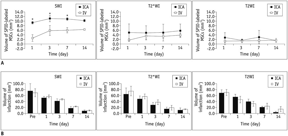
Fig. 8 Volumetric analysis of superparamagnetic iron oxide (SPIO)-labeled human bone marrow-derived mesenchymal stem cells (hBM-MSCs) and cerebral infarction. A. Volumes of engrafted SPIO-labeled hBM-MSCs in internal carotid arterial (ICA) group were larger than those in intravenous (IV) group on days 1, 3, and 7 (not on day 14) after injection on susceptibility-weighted images (SWI) (p < 0.05), but not on T2*-weighted images (T2*WI) and T2-weighted images (T2WI) (p > 0.05). SWI was most sensitive in detecting SPIO-labeled cells among MRI pulse sequences (p < 0.05). B. Volume of infarction decreased as time progressed, i.e., from day 2 after photothrombotic cerebral infarction (1 day before cell injection) until day 14 after stem cell injection in all 6 rats. Between ICA and IV groups, there were no differences in volume of cerebral infarction (p > 0.05).
Reference
-
1. Yang Y, Zhang J, Qian Y, Dong S, Huang H, Boada FE, et al. Superparamagnetic iron oxide is suitable to label tendon stem cells and track them in vivo with MR imaging. Ann Biomed Eng. 2013; 41:2109–2119.2. Bulte JW. In vivo MRI cell tracking: clinical studies. AJR Am J Roentgenol. 2009; 193:314–325.3. Richards JM, Shaw CA, Lang NN, Williams MC, Semple SI, MacGillivray TJ, et al. In vivo mononuclear cell tracking using superparamagnetic particles of iron oxide: feasibility and safety in humans. Circ Cardiovasc Imaging. 2012; 5:509–517.4. Hu SL, Lu PG, Zhang LJ, Li F, Chen Z, Wu N, et al. In vivo magnetic resonance imaging tracking of SPIO-labeled human umbilical cord mesenchymal stem cells. J Cell Biochem. 2012; 113:1005–1012.5. Reddy AM, Kwak BK, Shim HJ, Ahn C, Cho SH, Kim BJ, et al. Functional characterization of mesenchymal stem cells labeled with a novel PVP-coated superparamagnetic iron oxide. Contrast Media Mol Imaging. 2009; 4:118–126.6. Wang L, Deng J, Wang J, Xiang B, Yang T, Gruwel M, et al. Superparamagnetic iron oxide does not affect the viability and function of adipose-derived stem cells, and superparamagnetic iron oxide-enhanced magnetic resonance imaging identifies viable cells. Magn Reson Imaging. 2009; 27:108–119.7. Byun JS, Kwak BK, Kim JK, Jung J, Ha BC, Park S. Engraftment of human mesenchymal stem cells in a rat photothrombotic cerebral infarction model: comparison of intra-arterial and intravenous infusion using MRI and histological analysis. J Korean Neurosurg Soc. 2013; 54:467–476.8. Ha BC, Jung J, Kwak BK. Susceptibility-weighted imaging for stem cell visualization in a rat photothrombotic cerebral infarction model. Acta Radiol. 2015; 56:219–227.9. Xu HS, Ma C, Cao L, Wang JJ, Fan XX. Study of co-transplantation of SPIO labeled bone marrow stromal stem cells and Schwann cells for treating traumatic brain injury in rats and in vivo tracing of magnetically labeled cells by MRI. Eur Rev Med Pharmacol Sci. 2014; 18:520–525.10. Detante O, Valable S, de Fraipont F, Grillon E, Barbier EL, Moisan A, et al. Magnetic resonance imaging and fluorescence labeling of clinical-grade mesenchymal stem cells without impacting their phenotype: study in a rat model of stroke. Stem Cells Transl Med. 2012; 1:333–341.11. Vasconcelos-dos-Santos A, Rosado-de-Castro PH, Lopes de Souza SA, da Costa Silva J, Ramos AB, Rodriguez de Freitas G, et al. Intravenous and intra-arterial administration of bone marrow mononuclear cells after focal cerebral ischemia: is there a difference in biodistribution and efficacy? Stem Cell Res. 2012; 9:1–8.12. Walczak P, Zhang J, Gilad AA, Kedziorek DA, Ruiz-Cabello J, Young RG, et al. Dual-modality monitoring of targeted intraarterial delivery of mesenchymal stem cells after transient ischemia. Stroke. 2008; 39:1569–1574.13. Johnson GA, Calabrese E, Badea A, Paxinos G, Watson C. A multidimensional magnetic resonance histology atlas of the Wistar rat brain. Neuroimage. 2012; 62:1848–1856.14. Seki F, Hikishima K, Nambu S, Okanoya K, Okano HJ, Sasaki E, et al. Multidimensional MRI-CT atlas of the naked mole-rat brain (Heterocephalus glaber). Front Neuroanat. 2013; 7:45.15. Daadi MM, Li Z, Arac A, Grueter BA, Sofilos M, Malenka RC, et al. Molecular and magnetic resonance imaging of human embryonic stem cell-derived neural stem cell grafts in ischemic rat brain. Mol Ther. 2009; 17:1282–1291.16. Jung J, Kwak BK, Reddy AM, Ha BC, Shim HJ, Byun JS, et al. Characterization of photothrombotic cerebral infarction model at sensorimotor area of functional map in rat. J Neurol Sci-Turk. 2013; 30:617–628.17. Shen LH, Li Y, Chen J, Zhang J, Vanguri P, Borneman J, et al. Intracarotid transplantation of bone marrow stromal cells increases axon-myelin remodeling after stroke. Neuroscience. 2006; 137:393–399.18. Chen J, Zhang ZG, Li Y, Wang L, Xu YX, Gautam SC, et al. Intravenous administration of human bone marrow stromal cells induces angiogenesis in the ischemic boundary zone after stroke in rats. Circ Res. 2003; 92:692–699.19. Guzman R, Choi R, Gera A, De Los Angeles A, Andres RH, Steinberg GK. Intravascular cell replacement therapy for stroke. Neurosurg Focus. 2008; 24:E15.20. Pendharkar AV, Chua JY, Andres RH, Wang N, Gaeta X, Wang H, et al. Biodistribution of neural stem cells after intravascular therapy for hypoxic-ischemia. Stroke. 2010; 41:2064–2070.21. Lundberg J, Le Blanc K, Söderman M, Andersson T, Holmin S. Endovascular transplantation of stem cells to the injured rat CNS. Neuroradiology. 2009; 51:661–667.22. Li L, Jiang Q, Ding G, Zhang L, Zhang ZG, Li Q, et al. Effects of administration route on migration and distribution of neural progenitor cells transplanted into rats with focal cerebral ischemia, an MRI study. J Cereb Blood Flow Metab. 2010; 30:653–662.23. Fischer UM, Harting MT, Jimenez F, Monzon-Posadas WO, Xue H, Savitz SI, et al. Pulmonary passage is a major obstacle for intravenous stem cell delivery: the pulmonary first-pass effect. Stem Cells Dev. 2009; 18:683–692.24. Parr AM, Tator CH, Keating A. Bone marrow-derived mesenchymal stromal cells for the repair of central nervous system injury. Bone Marrow Transplant. 2007; 40:609–619.25. Cheng JL, Yang YJ, Li HL, Wang J, Wang MH, Zhang Y. In vivo tracing of superparamagnetic iron oxide-labeled bone marrow mesenchymal stem cells transplanted for traumatic brain injury by susceptibility weighted imaging in a rat model. Chin J Traumatol. 2010; 13:173–177.26. Jülke H, Veit C, Ribitsch I, Brehm W, Ludewig E, Delling U. Comparative labelling of equine and ovine multipotent stromal cells with superparamagnetic iron oxide particles for magnetic resonance imaging in vitro. Cell Transplant. 2013; 12. 10. [Epub].27. Pawelczyk E, Arbab AS, Chaudhry A, Balakumaran A, Robey PG, Frank JA. In vitro model of bromodeoxyuridine or iron oxide nanoparticle uptake by activated macrophages from labeled stem cells: implications for cellular therapy. Stem Cells. 2008; 26:1366–1375.28. Cohen ME, Muja N, Fainstein N, Bulte JW, Ben-Hur T. Conserved fate and function of ferumoxides-labeled neural precursor cells in vitro and in vivo. J Neurosci Res. 2010; 88:936–944.
- Full Text Links
-
- Actions
-
Cited
- CITED
-
- Close
- Share
-
- Similar articles
-
- Engraftment of Human Mesenchymal Stem Cells in a Rat Photothrombotic Cerebral Infarction Model : Comparison of Intra-Arterial and Intravenous Infusion Using MRI and Histological Analysis
- Comparison of Superparamagnetic Iron Oxide Labeling Efficiency between Poly-L-Lysine and Protamine Sulfate for Human Mesenchymal Stem Cells: Quantitative Analysis Using Multi-Echo T2* Magnetic Resonance Imaging
- In vivo Tracking of Mesenchymal Stem Cells Labeled with a Novel Chitosan-coated Superparamagnetic Iron Oxide Nanoparticles using 3.0T MRI
- Effects of Endothelial Progenitor Cells Used for Autograft Transplantation in Acute Myocardial Infarction Pig Model
- Monitoring Transplanted Human Mesenchymal Stem Cells in the Penile Cavernosal Tissues of Streptozotocin-induced Diabetic Rats Using Molecular Magnetic Resonance Imaging
