A Rare Case of Diffuse Pachymeningeal Involvement of Multiple Myeloma
- Affiliations
-
- 1Department of Radiology, Kyung Hee University Hospital, Seoul, Korea. euijkim@hanmail.net
- 2Department of Neurosurgery, Kyung Hee University Hospital, Seoul, Korea.
- KMID: 2151774
- DOI: http://doi.org/10.13104/imri.2015.19.4.252
Abstract
- Intracranial involvement in multiple myeloma patients takes up around 1%, and is usually known to be present in the parietal bone or skull base in cases of skull vault involvement, while it presents in the dura and parenchyma in cases of intracranial involvement. Primary pachymeningeal invasion is even rarer with extremely rapid progression and very poor prognosis. It is our intent to report a case in which we had to differentiate multiple myeloma with other metastatic tumors, lymphoma, and leukemia with intracranial involvement. Our patient showed an osteolytic lesion of the skull with dural involvement and subdural mass formations.
Keyword
Figure
-

Fig. 1 Whole spine MR and CT images of a 53-year-old female patient. The spine MR images (a-c) show diffuse heterogeneous signal intensity of vertebral bodies on both T1WI and T2WI. Homogeneously enhancing posterior epidural masses at the level of T4-5 and T10-12 also noted on the contrast enhanced T1WI (c). The whole spine CT image shows no evidence of bony destructive changes (d).

Fig. 2 Non-enhanced brain CT images of the patient. Multifocal osteolytic lesions of the skull bones (a) are noted. Also, a diffuse and high attenuated subdural lesion at left fronto-parietal convexity is present (b).
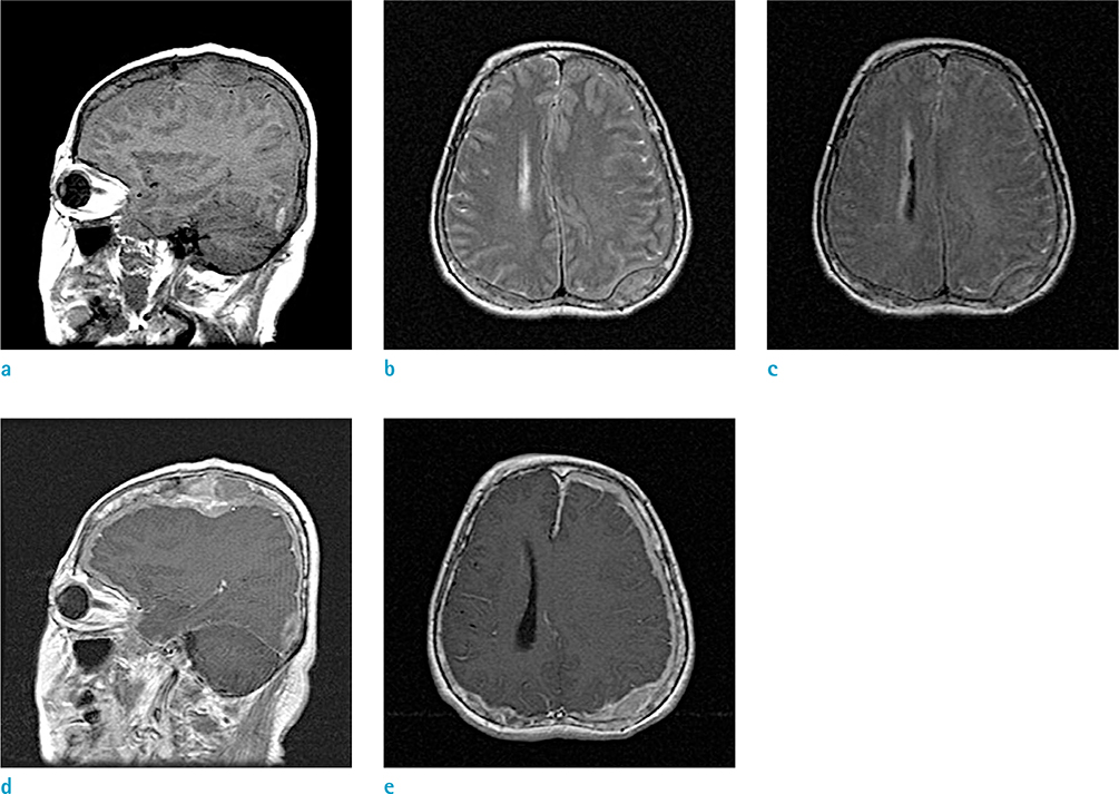
Fig. 3 Contrast enhanced brain MR images of the patient. The subdural mass shows iso signal intensity as the cerebral cortex on T1WI (a), T2WI (b), and FLAIR images (c). Diffuse heterogeneous enhancement of the mass along the thickened dura mater is noted on contrast enhanced T1WI (d, e).
Reference
-
1. Terada T. Multiple myeloma presenting as an intracranial plasmacytoma: a case report. Cases J. 2009; 2:9110.2. Cerase A, Tarantino A, Gozzetti A, et al. Intracranial involvement in plasmacytomas and multiple myeloma: a pictorial essay. Neuroradiology. 2008; 50:665–674.3. Munshi NC, Longo DL, Anderson KC. Plasma cell disorders. In : Longo DL, Fauci AS, Kasper DL, editors. Harrison's principles of internal medicine. 18th ed. New York: The McGraw-Hill Education;2011. p. 936–944.4. Schluterman KO, Fassas AB, Van Hemert RL, Harik SI. Multiple myeloma invasion of the central nervous system. Arch Neurol. 2004; 61:1423–1429.5. Pizzuti P, Pertuiset E, Chaumonnot F, et al. Neuromeningeal sites of multiple myeloma: 3 cases and review of the literature. Rev Med Interne. 1997; 18:646–651.6. De Blay V, Misson N, Dardenne G, Dupuis MJ. Leptomeningeal myelomatosis mimicking a subdural haematoma. Neuroradiology. 2000; 42:735–737.7. Roddie P, Collie D, Johnson P. Myelomatous involvement of the dura mater: a rare complication of multiple myeloma. J Clin Pathol. 2000; 53:398–399.8. Moran CC, Anderson CC, Caldemeyer KS, Smith RR. Meningeal myelomatosis: CT and MR appearances. AJNR Am J Neuroradiol. 1995; 16:1501–1503.
- Full Text Links
-
- Actions
-
Cited
- CITED
-
- Close
- Share
-
- Similar articles
-
- Liver Involvement of Multiple Myeloma Mimicking Intrahepatic Cholangiocarcinoma: A Case Report
- A Case of Diffuse Normolipemic Plane Xanthoma Associated with Multiple Myeloma
- Meningeal and Cerebral Involvement of a Plasmacytoma in an IgG Multiple Myeloma Patient: Case Report
- Diffuse Normolipemic Plane Xanthoma with Multiple Myeloma
- A Case of Diffuse Normolipemic Plane Xanthoma Associated with Multiple Myeloma
