Investig Magn Reson Imaging.
2015 Dec;19(4):231-236. 10.13104/imri.2015.19.4.231.
Tumor-like Presentation of Tubercular Brain Abscess: Case Report
- Affiliations
-
- 1Department of Radio-diagnosis, Patan Academy of Health Sciences, Patan, Nepal. kedibi@yahoo.com
- 2Department of Radio-diagnosis, Tribhuvan University Teaching Hospital, Kathmandu, Nepal.
- 3Department of Neurosurgery, Tribhuvan University Teaching Hospital, Kathmandu, Nepal.
- 4Department of Pathology, Tribhuvan University Teaching Hospital, Kathmandu, Nepal.
- 5Department of Neurology, National Academy of Medical Sciences, Bir Hospital, Kathmandu, Nepal.
- KMID: 2151770
- DOI: http://doi.org/10.13104/imri.2015.19.4.231
Abstract
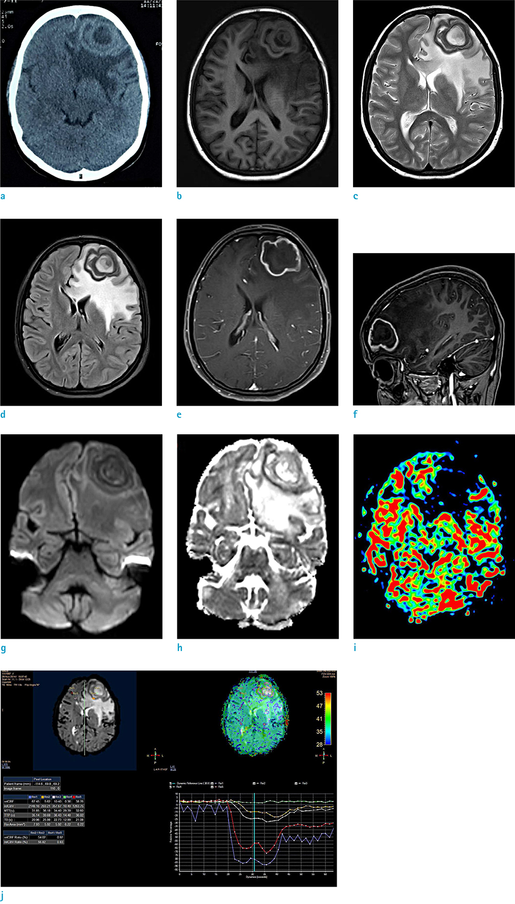
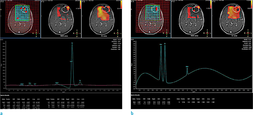
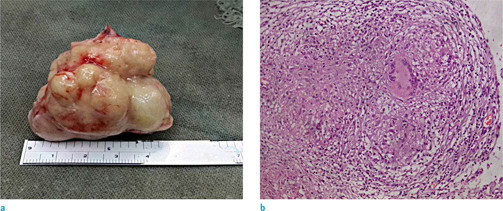
- A 17-year-old girl presented with complaints of headache and decreasing vision of one month's duration, without any history of fever, weight loss, or any evidence of an immuno-compromised state. Her neurological examination was normal, except for papilledema. Laboratory investigations were within normal limits, except for a slightly increased Erythrocyte Sedimentation Rate (ESR). Non-contrast computerized tomography of her head revealed complex mass in left frontal lobe with a concentric, slightly hyperdense, thickened wall, and moderate perilesional edema with mass effect. Differential diagnoses considered in this case were pilocytic astrocytoma, metastasis and abscess. Magnetic resonance imaging (MRI) obtained in 3.0 Tesla (3.0T) scanner revealed a lobulated outline cystic mass in the left frontal lobe with two concentric layers of T2 hypointense wall, with T2 hyperintensity between the concentric ring. Moderate perilesional edema and mass effect were seen. Post gadolinium study showed a markedly enhancing irregular wall with some enhancing nodular solid component. No restricted diffusion was seen in this mass in diffusion weighted imaging (DWI). Magnetic resonance spectroscopy (MRS) showed increased lactate and lipid peaks in the central part of this mass, although some areas at the wall and perilesional T2 hyperintensity showed an increased choline peak without significant decrease in N-acetylaspartate (NAA) level. Arterial spin labelling (ASL) and dynamic susceptibility contrast (DSC) enhanced perfusion study showed decrease in relative cerebral blood volume at this region. These features in MRI were suggestive of brain abscess. The patient underwent craniotomy with excision of a grayish nodular lesion. Abundant acid fast bacilli (AFB) in acid fast staining, and epithelioid cell granulomas, caseation necrosis and Langhans giant cells in histopathology, were conclusive of tubercular abscess. Tubercular brain abscess is a rare manifestation that simulates malignancy and cause diagnostic dilemma. MRI along with MRS and magnetic resonance perfusion studies, are powerful tools to differentiate lesions in such equivocal cases.
Keyword
MeSH Terms
-
Abscess
Adolescent
Astrocytoma
Blood Sedimentation
Blood Volume
Brain Abscess*
Brain*
Choline
Craniotomy
Diagnosis, Differential
Diffusion
Edema
Epithelioid Cells
Female
Fever
Frontal Lobe
Gadolinium
Giant Cells, Langhans
Granuloma
Head
Headache
Humans
Lactic Acid
Magnetic Resonance Imaging
Magnetic Resonance Spectroscopy
Necrosis
Neoplasm Metastasis
Neurologic Examination
Papilledema
Perfusion
Perfusion Imaging
Weight Loss
Choline
Gadolinium
Lactic Acid
Figure
Reference
-
1. National Tuberculosis Center. TB Burden in Nepal. Ministry of Health & Population;Accessed March 8, 2015. http://www.nepalntp.gov.np/.2. Cherian A, Thomas SV. Central nervous system tuberculosis. Afr Health Sci. 2011; 11:116–127.3. Jinkins JR, Gupta R, Chang KH, Rodriguez-Carbajal J. MR imaging of central nervous system tuberculosis. Radiol Clin North Am. 1995; 33:771–786.4. Trivedi R, Saksena S, Gupta RK. Magnetic resonance imaging in central nervous system tuberculosis. Indian J Radiol Imaging. 2009; 19:256–265.5. Bathla G, Khandelwal G, Maller VG, Gupta A. Manifestations of cerebral tuberculosis. Singapore Med J. 2011; 52:124–130. quiz 1316. Kastrup O, Wanke I, Maschke M. Neuroimaging of infections. NeuroRx. 2005; 2:324–332.7. Hansman Whiteman ML, Bowen BC, Donovan Post MJ, Bell MD. Intracranial infection. In : Atlas SW, editor. Magnetic resonance imaging of the brain and spine. 3rd ed. Philadelphia: Lippincott Williams & Wilkins;2002. p. 1099–1175.8. Desprechins B, Stadnik T, Koerts G, Shabana W, Breucq C, Osteaux M. Use of diffusion-weighted MR imaging in differential diagnosis between intracerebral necrotic tumors and cerebral abscesses. AJNR Am J Neuroradiol. 1999; 20:1252–1257.9. Holmes TM, Petrella JR, Provenzale JM. Distinction between cerebral abscesses and high-grade neoplasms by dynamic susceptibility contrast perfusion MRI. AJR Am J Roentgenol. 2004; 183:1247–1252.10. Luthra G, Parihar A, Nath K, et al. Comparative evaluation of fungal, tubercular, and pyogenic brain abscesses with conventional and diffusion MR imaging and proton MR spectroscopy. AJNR Am J Neuroradiol. 2007; 28:1332–1338.11. Mishra AM, Gupta RK, Jaggi RS, et al. Role of diffusion-weighted imaging and in vivo proton magnetic resonance spectroscopy in the differential diagnosis of ring-enhancing intracranial cystic mass lesions. J Comput Assist Tomogr. 2004; 28:540–547.12. Mishra AM, Gupta RK, Saksena S, et al. Biological correlates of diffusivity in brain abscess. Magn Reson Med. 2005; 54:878–885.13. Floriano VH, Ferraz-Filho JRL, Ronaldo Spotti A, Tognola WA. Perfusion-weighted magnetic resonance imaging in the evaluation of focal neoplastic and infectious brain lesions. Rev Bras Neurol. 2010; 46:29–36.14. Essig M, Nguyen TB, Shiroishi MS, et al. Perfusion MRI: the five most frequently asked clinical questions. AJR Am J Roentgenol. 2013; 201:W495–W510.15. Detre JA, Rao H, Wang DJ, Chen YF, Wang Z. Applications of arterial spin labeled MRI in the brain. J Magn Reson Imaging. 2012; 35:1026–1037.
- Full Text Links
- Actions
-
Cited
- CITED
-
- Close
- Share
- Similar articles
-
- Isolated Spontaneous Primary Tubercular Erector Spinae Abscess: A Case Report and Review of Literature
- A Case of Frontal Sinus Osteoma Causing Brain Abscess
- Brain Abscess Associated with Pulmonary Arteriovenous Malformation: Case Report
- Brain Abscess Following Intracerebral Hemorrhage: A Case Report
- Brain Abscess Diagnosed by Computerized Tomography: Case Report


