Extraneural Metastases of Glioblastoma without Simultaneous Central Nervous System Recurrence
- Affiliations
-
- 1Department of Neurosurgery, Seoul National University Hospital, Seoul, Korea.
- 2Neuro-Oncology Clinic, Research Institute and Hospital, National Cancer Center, Goyang, Korea. nslsh@ncc.re.kr
- KMID: 2134284
- DOI: http://doi.org/10.14791/btrt.2014.2.2.124
Abstract
- Glioblastoma multiforme (GBM) is well known as the most common malignant primary brain tumor. It could easily spread into the adjacent or distant brain tissue by infiltration, direct extension and cerebro-spinal fluid dissemination. The extranueural metastatic spread of GBM is relatively rare but it could have more progressive disease course. We report a 39-year-old man who had multiple bone metastases and malignant pleural effusion of the GBM without primary site recurrence.
MeSH Terms
Figure
-
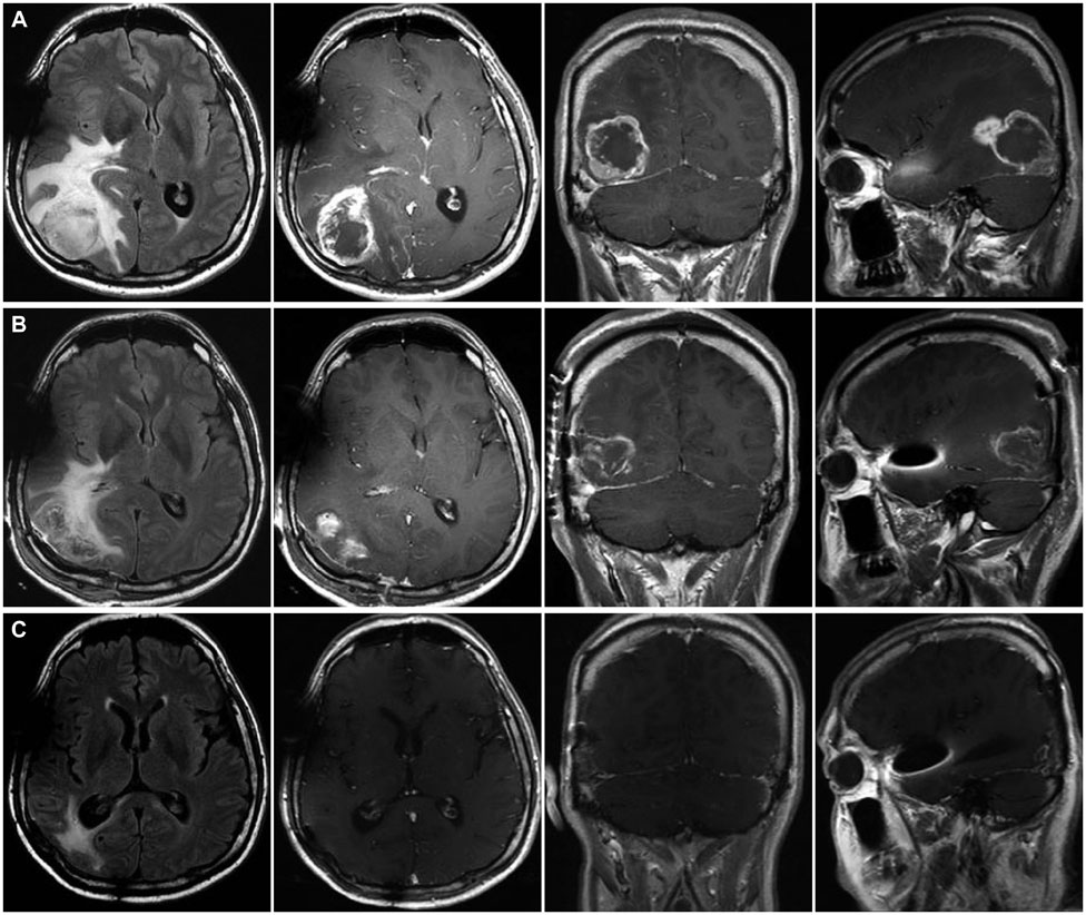
Fig. 1 A: Preoperative magnetic resonance imaging (MRI) reveals huge multi-lobulated contoured necrotic enhancing mass in right occipital lobe with extensive peritumoral edema. B: Postoperative MRI after one day-most of the enhancing mass are removed. C: Follow-up MRI at 10 months after surgery shows no evidence of tumor recurrence.
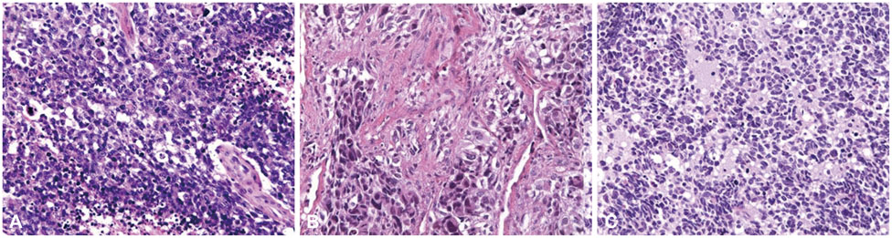
Fig. 2 Histologic findings of tumor mass from brain (A), spine (B), and rib (C) show pleomorphic astrocytic tumor cells with mitosis and nuclear atypia compatible to glioblastoma (hematoxylin-eosin, original magnification, ×400).
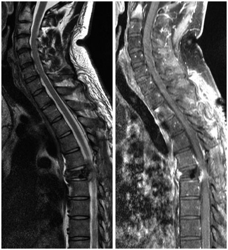
Fig. 3 Spine magnetic resonance imaging at 7 months after initial surgery reveals compressive pathologic fracture at the 6th thoracic vertebral body.
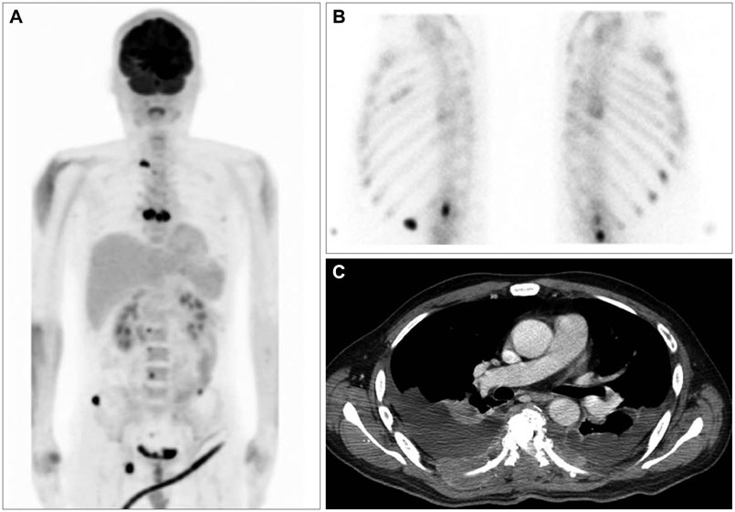
Fig. 4 A: Whole body fluorodeoxyglucose positron emission tomography scan imaging reveals multiple bone metastases to spine, ribs, pelvic, and scrum. B: Bone scan imaging shows multiple bone metastases in T-L spine and both ribs. C: Chest CT image reveals both malignant pleural effusion and both plural nodularity with focal chest wall invasion.
Reference
-
1. Shah A, Redhu R, Nadkarni T, Goel A. Supratentorial glioblastoma multiforme with spinal metastases. J Craniovertebr Junction Spine. 2010; 1:126–129.
Article2. Vertosick FT Jr, Selker RG. Brain stem and spinal metastases of supratentorial glioblastoma multiforme: a clinical series. Neurosurgery. 1990; 27:516–521. discussion 521-2.
Article3. Hsu E, Keene D, Ventureyra E, et al. Bone marrow metastasis in astrocytic gliomata. J Neurooncol. 1998; 37:285–293.4. Piccirilli M, Brunetto GM, Rocchi G, Giangaspero F, Salvati M. Extra central nervous system metastases from cerebral glioblastoma multiforme in elderly patients. Clinico-pathological remarks on our series of seven cases and critical review of the literature. Tumori. 2008; 94:40–51.
Article5. Rajagopalan V, El Kamar FG, Thayaparan R, Grossbard ML. Bone marrow metastases from glioblastoma multiforme--a case report and review of the literature. J Neurooncol. 2005; 72:157–161.
Article6. Mentrikoski M, Johnson MD, Korones DN, Scott GA. Glioblastoma multiforme in skin: a report of 2 cases and review of the literature. Am J Dermatopathol. 2008; 30:381–384.
Article7. Bernstein JJ, Woodard CA. Glioblastoma cells do not intravasate into blood vessels. Neurosurgery. 1995; 36:124–132. discussion 132.
Article8. Huang P, Allam A, Taghian A, Freeman J, Duffy M, Suit HD. Growth and metastatic behavior of five human glioblastomas compared with nine other histological types of human tumor xenografts in SCID mice. J Neurosurg. 1995; 83:308–315.
Article
- Full Text Links
-
- Actions
-
Cited
- CITED
-
- Close
- Share
-
- Similar articles
-
- Extraneural Metastasis of Glioblastoma Multiforme Presenting as an Unusual Neck Mass
- Clinical Analysis of 47 Cases of Intracranial Glioma
- Radiation-Induced Glioblastoma Multiforme in a Remitted Acute Lymphocytic Leukemia Patient
- The value of computerized axial tomography of the brain in children with central nervous system disorders
- Bilateral Triple-Negative Invasive Breast Cancer with a BRCA2 Mutation, and Glioblastoma: A Case Report and Literature Review
