J Korean Soc Magn Reson Med.
2012 Aug;16(2):115-123. 10.13104/jksmrm.2012.16.2.115.
Evaluation of White Matter Abnormality in Mild Alzheimer Disease and Mild Cognitive Impairment Using Diffusion Tensor Imaging: A Comparison of Tract-Based Spatial Statistics with Voxel-Based Morphometry
- Affiliations
-
- 1Department of Radiology and Research Institute of Radiology, University of Ulsan College of Medicine, Asan Medical Center, Korea. sjkimjb@amc.seoul.kr
- 2Department of Neurology, University of Ulsan College of Medicine, Asan Medical Center, Korea.
- 3Department of Psychiatry and Health Promotion Center, University of Ulsan College of Medicine, Asan Medical Center, Korea.
- 4Division of Magnetic Resonance, Korea Basic Science Institute, Ochang-Eup, Korea.
- 5Department of Radiology, Kyung Hee East-West Neo Medial Center, Kyung Hee University College of Medicine, Seoul, Korea.
- KMID: 2099845
- DOI: http://doi.org/10.13104/jksmrm.2012.16.2.115
Abstract
- PURPOSE
To evaluate white matter abnormalities on diffusion tensor imaging (DTI) in patients with mild Alzheimer disease (AD) and mild cognitive impairment (MCI), using tract-based spatial statistics (TBSS) and voxel-based morphometry (VBM).
MATERIALS AND METHODS
DTI was performed in 21 patients with mild AD, in 13 with MCI and in 16 old healthy subjects. A fractional anisotropy (FA) map was generated for each participant and processed for voxel-based comparisons among the three groups using TBSS. For comparison, DTI data was processed using the VBM method, also.
RESULTS
TBSS showed that FA was significantly lower in the AD than in the old healthy group in the bilateral anterior and right posterior corona radiata, the posterior thalamic radiation, the right superior longitudinal fasciculus, the body of the corpus callosum, and the right precuneus gyrus. VBM identified additional areas of reduced FA, including both uncinates, the left parahippocampal white matter, and the right cingulum. There were no significant differences in FA between the AD and MCI groups, or between the MCI and old healthy groups.
CONCLUSION
TBSS showed multifocal abnormalities in white matter integrity in patients with AD compared with old healthy group. VBM could detect more white matter lesions than TBSS, but with increased artifacts.
Keyword
MeSH Terms
Figure
-
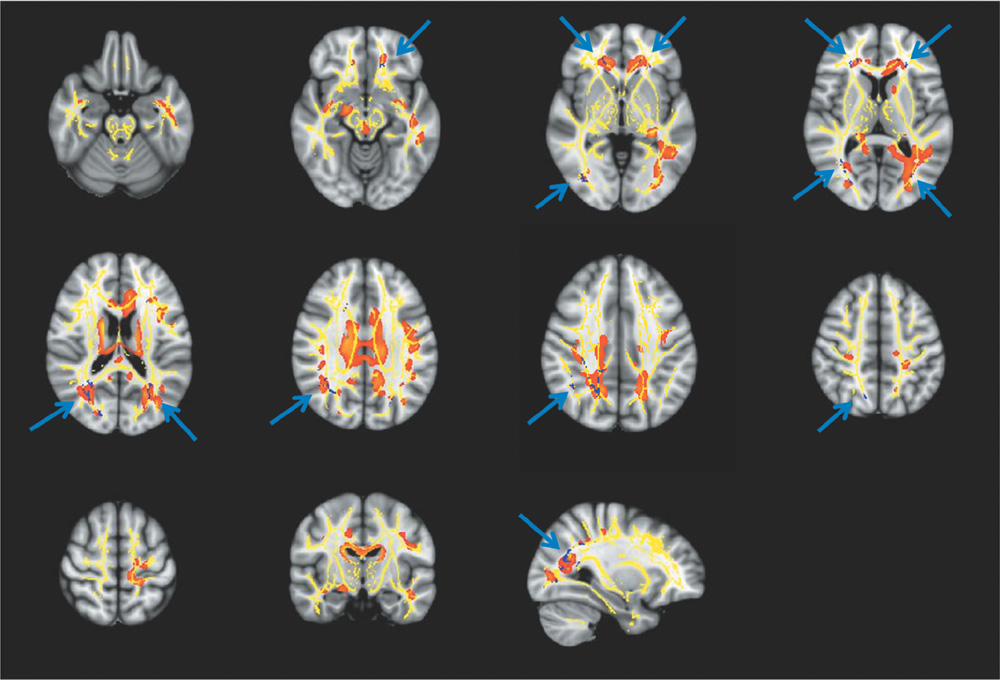
Fig. 1 Results of TBSS and VBM analyses of patients with mild AD. The figure shows the patterns of reduced FA in mild AD subjects (blue) compared with old healty subjects at TBSS analysis, overlaid onto a mean FA skeleton (yellow). The area of reduced FA (in red) at VBM analysis is also shown (p < 0.05, corrected for multiple comparisons). Note.- TBSS = tract-based spatial statistics; VBM = voxel-based morphometry; AD = Alzheimer disease; FA = fractional anisotrophy
Reference
-
1. Petersen RC, Doody R, Kurz A, et al. Current concepts in mild cognitive impairment. Arch Neurol. 2001. 58:1985–1992.2. Leifer D, Buonanno FS, Richardson EP. Clinicopathologic correlations of cranial magnetic resonance imaging of periventricular white matter. Neurology. 1990. 40:911–918.3. de Leeuw FE, Barkhof F, Scheltens P. White matter lesions and hippocampal atrophy in Alzheimer's disease. Neurology. 2004. 62:310–312.4. Bozzali M, Falini A, Franceschi M, et al. White matter damage in Alzheimer's disease assessed in vivo using diffusion tensor magnetic resonance imaging. J Neurol Neurosurg Psychiatry. 2002. 72:742–746.5. Sydykova D, Stahl R, Dietrich O, et al. Fiber connections between the cerebral cortex and the corpus callosum in Alzheimer's disease: a diffusion tensor imaging and voxel-based morphometry study. Cereb Cortex. 2007. 17:2276–2282.6. Yoshita M, Fletcher E, Harvey D, et al. Extent and distribution of white matter hyperintensities in normal aging, MCI, and AD. Neurology. 2006. 67:2192–2198.7. Serra L, Cercignani M, Lenzi D, et al. Grey and white matter changes at different stages of Alzheimer's disease. J Alzheimers Dis. 2010. 19:147–159.8. Chetelat G, Desgranges B, Landeau B, et al. Direct voxel-based comparison between grey matter hypometabolism and atrophy in Alzheimer's disease. Brain. 2008. 131:60–71.9. Xie S, Xiao JX, Gong GL, et al. Voxel-based detection of white matter abnormalities in mild Alzheimer disease. Neurology. 2006. 66:1845–1849.10. Medina D, DeToledo-Morrell L, Urresta F, et al. White matter changes in mild cognitive impairment and AD: a diffusion tensor imaging study. Neurobiol Aging. 2006. 27:663–672.11. Stahl R, Dietrich O, Teipel SJ, Hampel H, Reiser MF, Schoenberg SO. White matter damage in Alzheimer disease and mild cognitive impairment: assessment with diffusion-tensor MR imaging and parallel imaging techniques. Radiology. 2007. 243:483–492.12. Takahashi S, Yonezawa H, Takahashi J, Kudo M, Inoue T, Tohgi H. Selective reduction of diffusion anisotropy in white matter of Alzheimer disease brains measured by 3.0 Tesla magnetic resonance imaging. Neurosci Lett. 2002. 332:45–48.13. Salat DH, Greve DN, Pacheco JL, et al. Regional white matter volume differences in nondemented aging and Alzheimer's disease. Neuroimage. 2009. 44:1247–1258.14. Stoub TR, deToledo-Morrell L, Stebbins GT, Leurgans S, Bennett DA, Shah RC. Hippocampal disconnection contributes to memory dysfunction in individuals at risk for Alzheimer's disease. Proc Natl Acad Sci U S A. 2006. 103:10041–10045.15. Chaim TM, Duran FL, Uchida RR, Perico CA, de Castro CC, Busatto GF. Volumetric reduction of the corpus callosum in Alzheimer's disease in vivo as assessed with voxel-based morphometry. Psychiatry Res. 2007. 154:59–68.16. Smith SM, Jenkinson M, Johansen-Berg H, et al. Tract-based spatial statistics: voxelwise analysis of multi-subject diffusion data. Neuroimage. 2006. 31:1487–1505.17. McKhann G, Drachman D, Folstein M, Katzman R, Price D, Stadlan EM. Clinical diagnosis of Alzheimer's disease: report of the NINCDS-ADRDA work group under the auspices of department of health and human services task force on Alzheimer's disease. Neurology. 1984. 34:939–944.18. Folstein MF, Folstein SE, McHugh PR. "Mini-mental state". A practical method for grading the cognitive state of patients for the clinician. J Psychiatr Res. 1975. 12:189–198.19. Smith SM, Nichols TE. Threshold-free cluster enhancement: addressing problems of smoothing, threshold dependence and localisation in cluster inference. Neuroimage. 2009. 44:83–98.20. Wakana S, Jiang H, Nagae-Poetscher LM, van Zijl PC, Mori S. Fiber tract-based atlas of human white matter anatomy. Radiology. 2004. 230:77–87.21. Wakana S, Caprihan A, Panzenboeck MM, et al. Reproducibility of quantitative tractography methods applied to cerebral white matter. Neuroimage. 2007. 36:630–644.22. Mori S, Wakana S, van Zijl PCM, Nagae-Poetscher LM. MRI atlas of human white matter. 2005. Amsterdam, The Netherland: Elsevier;15–237.23. Bosch B, Arenaza-Urquijo EM, Rami L, et al. Multiple DTI index analysis in normal aging, amnestic MCI and AD. Relationship with neuropsychological performance. Neurobiol Aging. 2010. 04. 03. [Epub ahead of print]. doi: 10.1016/j.neurobiolaging.2010.02.004 .24. Liu Y, Spulber G, Lehtimaki KK, et al. Diffusion tensor imaging and tract-based spatial statistics in Alzheimer's disease and mild cognitive impairment. Neurobiol Aging. 2011. 32:1558–1571.25. Stricker NH, Schweinsburg BC, Delano-Wood L, et al. Decreased white matter integrity in late-myelinating fiber pathways in Alzheimer's disease supports retrogenesis. Neuroimage. 2009. 45:10–16.26. Balthazar ML, Yasuda CL, Pereira FR, Pedro T, Damasceno BP, Cendes F. Differences in grey and white matter atrophy in amnestic mild cognitive impairment and mild Alzheimer's disease. Eur J Neurol. 2009. 16:468–474.27. Zhuang L, Wen W, Zhu W, et al. White matter integrity in mild cognitive impairment: a tract-based spatial statistics study. Neuroimage. 2010. 53:16–25.28. Salat DH, Tuch DS, van der Kouwe AJ, et al. White matter pathology isolates the hippocampal formation in Alzheimer's disease. Neurobiol Aging. 2010. 31:244–256.29. Fellgiebel A, Scheurich A, Bartenstein P, Muller MJ. FDG-PET and CSF phospho-tau for prediction of cognitive decline in mild cognitive impairment. Psychiatry Res. 2007. 155:167–171.30. Acosta-Cabronero J, Williams GB, Pengas G, Nestor PJ. Absolute diffusivities define the landscape of white matter degeneration in Alzheimer's disease. Brain. 2010. 133:529–539.31. Smith CD, Chebrolu H, Andersen AH, et al. White matter diffusion alterations in normal women at risk of Alzheimer's disease. Neurobiol Aging. 2010. 31:1122–1131.32. Damoiseaux JS, Smith SM, Witter MP, et al. White matter tract integrity in aging and Alzheimer's disease. Hum Brain Mapp. 2009. 30:1051–1059.
- Full Text Links
-
- Actions
-
Cited
- CITED
-
- Close
- Share
-
- Similar articles
-
- Gray and White Matter Degenerations in Subjective Memory Impairment: Comparisons with Normal Controls and Mild Cognitive Impairment
- Topographical Disorientation in Mild Cognitive Impairment: A Voxel-Based Morphometry Study
- Development of a Korean Standard Structural Brain Template in Cognitive Normals and Patients with Mild Cognitive Impairment and Alzheimer's Disease
- Morphological and Microstructural Changes of the Hippocampus in Early MCI: A Study Utilizing the Alzheimer's Disease Neuroimaging Initiative Database
- Association between Cognitive Function, Behavioral and Psychological Symptoms of Dementia and White Matter Hyperintensities in Patients with Alzheimer's Disease and Mild Cognitive Impairment
