Topographical Disorientation in Mild Cognitive Impairment: A Voxel-Based Morphometry Study
- Affiliations
-
- 1Department of Neurology, Ajou University School of Medicine, Suwon, Korea. symoon.bv@gmail.com
- 2Department of Psychology, University of Calgary, Calgary, Canada.
- KMID: 1462808
- DOI: http://doi.org/10.3988/jcn.2010.6.4.204
Abstract
- BACKGROUND AND PURPOSE
To assess the neural substrates underlying topographical disorientation (TD) in patients affected by mild cognitive impairment (MCI), forty-one patients diagnosed with MCI and 24 healthy control individuals were recruited.
METHODS
TD was assessed clinically in all participants. Neurological and neuropsychological evaluations and a volumetric-head magnetic resonance imaging scan were performed in each participant. Voxel-based morphometry was used to compare patterns of gray-matter atrophy between patients with and without TD, and a group of normal controls.
RESULTS
We found TD in 17 out of the 41 MCI patients (41.4%). The functional abilities were significantly impaired in MCI patients with TD compared to in MCI patients without TD. Voxel-based morphometry analyses showed that the presence of TD in MCI patients is associated with loss of gray matter in the medial temporal regions, including the hippocampus and parahippocampal cortex, the fusiform gyrus, the inferior occipital gyrus, the amygdala, and the cerebellum.
CONCLUSIONS
The findings found in this study represent the first evidence that the presence of TD in patients with MCI is associated with loss of gray matter in those brain regions that have been documented to be responsible for orientation in both neuropsychological and neuroimaging studies.
MeSH Terms
Figure
-
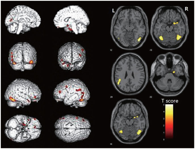
Fig. 1 Patterns of statistically significant loss of gray matter in patients with mild cognitive impairment (MCI) in combination with topographical disorientation (TD) compared to healthy control subjects (p<0.001, uncorrected for multiple comparisons). In the right hemisphere, MCI-TD patients exhibited loss of gray matter from the anterior temporal pole, through the medial temporal regions (including the parahippocampal and fusiform gyri), extending to the middle and inferior temporal gyri toward the most posterior regions of the fusiform and inferior occipital gyri. Similar regions were involved in the left hemisphere, although to a lesser extent. In addition, gray matter loss was observed in the right amygdala, left angular gyrus, and cerebellum.
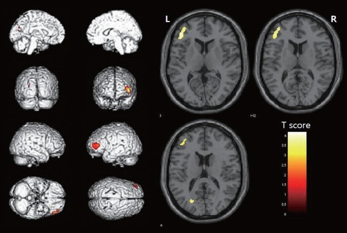
Fig. 2 Patterns of statistically significant loss of gray matter in patients with MCI without TD (MCI-noTD) compared to healthy control subjects (p<0.001, uncorrected for multiple comparisons). MCI-noTD patients exhibited gray matter loss restricted to the left frontal and occipital regions when compared to the group of healthy controls.
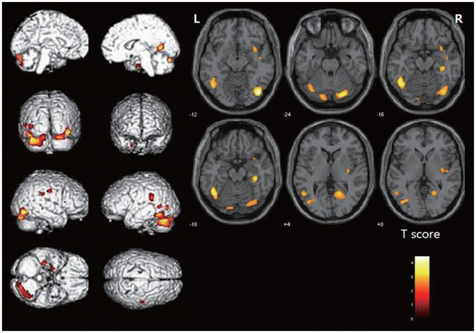
Fig. 3 Regions of gray matter loss in mild cognitive impairment-topographical disorientation (MCI-TD) patients compared to MCI-noTD patients (p<0.01, uncorrected for multiple comparisons). MCI-TD patients exhibited a greater loss of gray matter loss bilaterally in the temporo-occipital regions. These regions included the hippocampi and parahippocampal gyri, the fusiform gyrus, the inferior occipital gyrus, the amygdala, and the cerebellum.
Reference
-
1. Renzi ED. Disorders of space exploration and cognition. 1982. Chichester: Wiley.2. Aguirre GK, D'Esposito M. Topographical disorientation: a synthesis and taxonomy. Brain. 1999. 122:1613–1628.
Article3. Habib M, Sirigu A. Pure topographical disorientation: a definition and anatomical basis. Cortex. 1987. 23:73–85.
Article4. Pallis CA. Impaired identification of faces and places with agnosia for colours; report of a case due to cerebral embolism. J Neurol Neurosurg Psychiatry. 1955. 18:218–224.
Article5. Takahashi N, Kawamura M, Shiota J, Kasahata N, Hirayama K. Pure topographic disorientation due to right retrosplenial lesion. Neurology. 1997. 49:464–469.
Article6. Stark M. Impairment of an Egocentric Map of Locations: Implications for Perception and Action. Cogn Neuropsychol. 1996. 13:481–524.
Article7. Barrash J. A historical review of topographical disorientation and its neuroanatomical correlates. J Clin Exp Neuropsychol. 1998. 20:807–827.
Article8. Huang C, Wahlund LO, Svensson L, Winblad B, Julin P. Cingulate cortex hypoperfusion predicts Alzheimer's disease in mild cognitive impairment. BMC Neurol. 2002. 2:9.
Article9. Trivedi MA, Wichmann AK, Torgerson BM, Ward MA, Schmitz TW, Ries ML, et al. Structural MRI discriminates individuals with Mild Cognitive Impairment from age-matched controls: a combined neuropsychological and voxel based morphometry study. Alzheimers Dement. 2006. 2:296–302.
Article10. Seo SW, Im K, Lee JM, Kim YH, Kim ST, Kim SY, et al. Cortical thickness in single- versus multiple-domain amnestic mild cognitive impairment. Neuroimage. 2007. 36:289–297.
Article11. Desikan RS, Fischl B, Cabral HJ, Kemper TL, Guttmann CR, Blacker D, et al. MRI measures of temporoparietal regions show differential rates of atrophy during prodromal AD. Neurology. 2008. 71:819–825.
Article12. Whitwell JL, Przybelski SA, Weigand SD, Knopman DS, Boeve BF, Petersen RC, et al. 3D maps from multiple MRI illustrate changing atrophy patterns as subjects progress from mild cognitive impairment to Alzheimer's disease. Brain. 2007. 130:1777–1786.
Article13. Hort J, Laczó J, Vyhnálek M, Bojar M, Bures J, Vlcek K. Spatial navigation deficit in amnestic mild cognitive impairment. Proc Natl Acad Sci U S A. 2007. 104:4042–4047.
Article14. Petersen RC. Mild cognitive impairment as a diagnostic entity. J Intern Med. 2004. 256:183–194.
Article15. Kang Y, Na D. Seoul Neuropsychological Screening Battery. 2003. Incheon: Human Brain Research and Consulting.16. Cummings JL, Mega M, Gray K, Rosenberg-Thompson S, Carusi DA, Gornbein J. The Neuropsychiatric Inventory: comprehensive assessment of psychopathology in dementia. Neurology. 1994. 44:2308–2314.
Article17. Morris JC. The Clinical Dementia Rating (CDR): current version and scoring rules. Neurology. 1993. 43:2412–2414.18. Ku HM, Kim JH, Kwon EJ, Kim SH, Lee HS, Ko HJ, et al. A study on the reliability and validity of seoul-instrumental activities of daily living (S-IADL). J Korean Neuropsychiatr Assoc. 2004. 43:189–199.19. Pai MC, Jacobs WJ. Topographical disorientation in community-residing patients with Alzheimer's disease. Int J Geriatr Psychiatry. 2004. 19:250–255.
Article20. Kim H, Na D. Normative data on the Korean version of the Boston Naming Test. J Clin Exp Neuropsychol. 1999. 21:127–133.21. Ashburner J, Friston KJ. Voxel-based morphometry--the methods. Neuroimage. 2000. 11:805–821.
Article22. Senjem ML, Gunter JL, Shiung MM, Petersen RC, Jack CR Jr. Comparison of different methodological implementations of voxel-based morphometry in neurodegenerative disease. Neuroimage. 2005. 26:600–608.
Article23. Bozzali M, Filippi M, Magnani G, Cercignani M, Franceschi M, Schiatti E, et al. The contribution of voxel-based morphometry in staging patients with mild cognitive impairment. Neurology. 2006. 67:453–460.
Article24. Rondi-Reig L, Burguière E. Is the cerebellum ready for navigation? Prog Brain Res. 2005. 148:199–212.
Article25. Lee TM, Liu HL, Hung KN, Pu J, Ng YB, Mak AK, et al. The cerebellum's involvement in the judgment of spatial orientation: a functional magnetic resonance imaging study. Neuropsychologia. 2005. 43:1870–1877.
Article26. Burguière E, Arleo A, Hojjati M, Elgersma Y, De Zeeuw CI, Berthoz A, et al. Spatial navigation impairment in mice lacking cerebellar LTD: a motor adaptation deficit? Nat Neurosci. 2005. 8:1292–1294.
Article27. Aguirre GK, Zarahn E, D'Esposito M. An area within human ventral cortex sensitive to "building" stimuli: evidence and implications. Neuron. 1998. 21:373–383.
Article28. Maguire EA. Hippocampal involvement in human topographical memory: evidence from functional imaging. Philos Trans R Soc Lond B Biol Sci. 1997. 352:1475–1480.
Article29. Iaria G, Chen JK, Guariglia C, Ptito A, Petrides M. Retrosplenial and hippocampal brain regions in human navigation: complementary functional contributions to the formation and use of cognitive maps. Eur J Neurosci. 2007. 25:890–899.
Article30. Iaria G, Lanyon LJ, Fox CJ, Giaschi D, Barton JJ. Navigational skills correlate with hippocampal fractional anisotropy in humans. Hippocampus. 2008. 18:335–339.
Article31. Epstein RA, Parker WE, Feiler AM. Where am I now? Distinct roles for parahippocampal and retrosplenial cortices in place recognition. J Neurosci. 2007. 27:6141–6149.
Article32. Ekstrom AD, Kahana MJ, Caplan JB, Fields TA, Isham EA, Newman EL, et al. Cellular networks underlying human spatial navigation. Nature. 2003. 425:184–188.
Article33. Epstein R, Kanwisher N. A cortical representation of the local visual environment. Nature. 1998. 392:598–601.
Article34. Grön G, Wunderlich AP, Spitzer M, Tomczak R, Riepe MW. Brain activation during human navigation: gender-different neural networks as substrate of performance. Nat Neurosci. 2000. 3:404–408.
Article35. Zeki S. Cerebral akinetopsia (visual motion blindness). A review. Brain. 1991. 114:811–824.36. Jahn K, Deutschländer A, Stephan T, Strupp M, Wiesmann M, Brandt T. Brain activation patterns during imagined stance and locomotion in functional magnetic resonance imaging. Neuroimage. 2004. 22:1722–1731.
Article37. Chételat G, Desgranges B, De La Sayette V, Viader F, Eustache F, Baron JC. Mapping gray matter loss with voxel-based morphometry in mild cognitive impairment. Neuroreport. 2002. 13:1939–1943.
Article38. Karas GB, Scheltens P, Rombouts SA, Visser PJ, van Schijndel RA, Fox NC, et al. Global and local gray matter loss in mild cognitive impairment and Alzheimer's disease. Neuroimage. 2004. 23:708–716.
Article39. Pennanen C, Testa C, Laakso MP, Hallikainen M, Helkala EL, Hänninen T, et al. A voxel based morphometry study on mild cognitive impairment. J Neurol Neurosurg Psychiatry. 2005. 76:11–14.
Article40. Bell-McGinty S, Lopez OL, Meltzer CC, Scanlon JM, Whyte EM, Dekosky ST, et al. Differential cortical atrophy in subgroups of mild cognitive impairment. Arch Neurol. 2005. 62:1393–1397.
Article41. Fischer P, Jungwirth S, Zehetmayer S, Weissgram S, Hoenigschnabl S, Gelpi E, et al. Conversion from subtypes of mild cognitive impairment to Alzheimer dementia. Neurology. 2007. 68:288–291.
Article42. Money J, Alexander D, Walker H. Astandardized road-map test of direction sense. 1976. Baltimore, MD: Johns Hopkins University Press.43. Hunt LA, Murphy CF, Carr D, Ducherk JM, Buckles V, Morris JC. Environmental cueing may effect performance on a road test for drivers with dementia of the Alzheimer type. Alzheimer Dis Assoc Disord. 1997. 11:Suppl 1. 13–16.
Article44. Nadolne MJ, Stringer AY. Ecologic validity in neuropsychological assessment: prediction of wayfinding. J Int Neuropsychol Soc. 2001. 7:675–682.
Article
- Full Text Links
-
- Actions
-
Cited
- CITED
-
- Close
- Share
-
- Similar articles
-
- Erratum: Topographical Disorientation in Mild Cognitive Impairment: A Voxel-Based Morphometry Study
- Evaluation of White Matter Abnormality in Mild Alzheimer Disease and Mild Cognitive Impairment Using Diffusion Tensor Imaging: A Comparison of Tract-Based Spatial Statistics with Voxel-Based Morphometry
- Gray and White Matter Degenerations in Subjective Memory Impairment: Comparisons with Normal Controls and Mild Cognitive Impairment
- Impact of Apolipoprotein E ε4 in Alzheimer’s Disease: A Meta-Analysis of Voxel-Based Morphometry Studies
- Effects of Cognitive-based Interventions of Older Adults with Mild Cognitive Impairment: A Systematic Review and Meta-analysis
