Water Extract of Samultang Reduces Apoptotic Cell Death by H2O2-Induced Oxidative Injury in SK-N-MC Cells
- Affiliations
-
- 1Department of Physiology, Wonkwang University School of Medicine, Iksan 570-749, Korea. mskim@wku.ac.kr
- KMID: 2071663
- DOI: http://doi.org/10.4196/kjpp.2009.13.3.139
Abstract
- The purpose of this study was to evaluate the effects of the water extract of Samultang (SMT), a Chinese herb, on apoptotic cell death by H2O2-induced oxidative stress in SK-N-MC cells. A nuclear fragmentation was observed via fluorescence imaging 12 h after exposure to 30 micrometer H2O2 and DNA laddering was detected via agarose electrophoresis gel. In addition, increases in sub-G1 phase and cleavage of the PARP protein were observed. However, treatment with SMT for 2 h prior to H2O2 exposure significantly reduced apoptotic cell death induced by incubation with 30 micrometer H2O2 in SK-N-MC cells. Pre-incubation with water extract of SMT for 2 h prevented the H2O2-induced decrease in mitochondrial transmembrane potential. SMT also attenuated the increase in caspase-3 activity and the breakdown of PARP protein caused by H2O2-induced oxidative stress. These results suggest that the water extract of SMT provides inhibition of apoptotic cell death against oxidative injury in SK-N-MC cells.
Keyword
MeSH Terms
Figure
-

Fig. 1. Effects of H2O2 or SMT on cell viability in SK-N-MC cells. (A) Dose-response relationship of H2O2 on cell viability, (B) time-dependent relationship of H2O2 (30 μM) on cell viability, (C) dose-response relationship of SMT, (D) effect of SMT pretreatment on H2O2-induced cytotoxicity in SK-N-MC cells. The various concentrations of SMT were given 2 h before 30 μM H2O2 treatment. Cell viability was determined by MTT assay after 12 h H2O2 treatment. Values are mean±S.D. of each triplet (n=3). ∗Denotes a significant difference between H2O2 and H2O2 + SMT (∗p<0.05, ∗∗p<0.01).

Fig. 2. Photographs of DAPI staining showing changes in DNA morphology of SK-N-MC cells. (A) Control cells not treated with H2O2, (B) cells treated with 30 μM H2O2 for 12 h, (C) cells treated with SMT (300 μg/ml) 2 h prior to 30 μM H2O2, (D) bar histogram showing percent of cells showing abnormal nuclear morphology counted in 3 randomly selected fields. A large amount of DNA was condensed or fragmented in cells treated only with H2O2 (arrows), but pretreatment with SMT significantly reduced DNA abnormalities. Values are mean±S.D. of each triplet (n=3). ∗Denotes a significant difference between H2O2 and H2O2 + SMT (∗p<0.05).
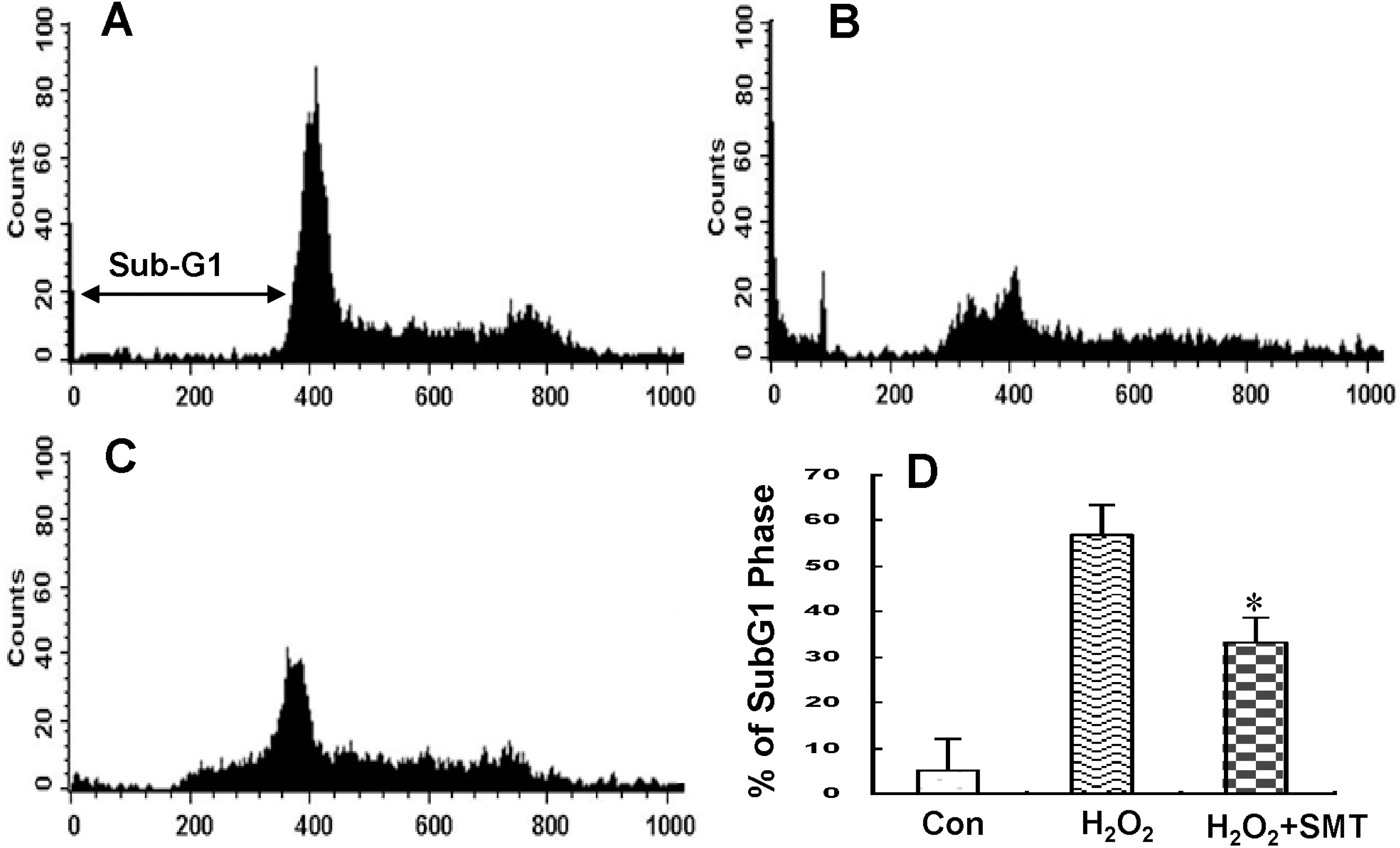
Fig. 3. Flow cytometric analysis showing changes in cell cycle of SK-N-MC cells. (A) Control, (B) treatment with 30 μM H2O2, (C) SMT (300 μg/ml) treatment 2 h prior to exposure to H2O2 for 12 h, (D) bar histogram showing percent changes of sub-G1 phase in cell cycle. Values are mean± S.D. of each triplet (n=3). ∗Denotes a significant difference between H2O2 and H2O2 + SMT (∗p<0.05).

Fig. 4. Agarose gel electrophoresis of DNA extracted from SK-N-MC cells. Lane A, DNA marker (100 bp); Lane B, control; Lane C, treatment with 30 μM H2O2 for 12 h; Lane D, SMT (300 μg/ml) treatment 2 h prior to exposure to H2O2 for 12 h.

Fig. 5. Flow cytometric analysis showing changes in the mitochondrial membrane potential of SK-N-MC cells. (A) Control, (B) treatment with 30 μM H2O2, (C) SMT (300 μg/ml) treatment 2 h prior to exposure to H2O2 for 12 h, (D) bar histogram showing the percent changes of relative fluorescence intensity (RFI) of mitochondrial membrane potential in control, H2O2-only treated, and SMT plus H2O2 treated SK-N-MC cells. Values are means±S.D. of each triplet (n=3). ∗Denotes a significant difference between H2O2 and H2O2 + SMT (∗p<0.05).

Fig. 6. RT-PCR analysis of caspase-3 mRNA expression from the cDNA of SK-N-MC cells 3 h after H2O2 treatment. Control, cells not treated with H2O2; H2O2, cells treated with 30 μM H2O2 for 3 h; SMT-H2O2, cells treated with 300 μg/ml SMT 2 h prior to 30 μM H2O2. Values are means±S.D. of each triplet (n=3). ∗Denotes a significant difference between H2O2 and H2O2 + SMT (∗p<0.05).

Fig. 7. Changes in caspase-3 enzyme activity from whole cell lysate of SK-N-MC cells. (A) Caspase-3 enzyme activity at 6 and 12 h after 30 μM H2O2 treatment, (B) effect of SMT (300 μg/ml) pretreatment on caspase-3 enzyme activity 12 h after H2O2 treatment. Values are means±S.D. of each triplet (n=3). ∗Denotes a significant difference between H2O2 and H2O2 + SMT (∗p<0.05, ∗∗p<0.01).

Fig. 8. Western blot analysis for expression of the PARP protein from whole cell lysates of SK-N-MC cells 12 h after H2O2 treatment. Control, cells not treated with H2O2; H2O2, cells treated with 30 μM H2O2 for 12 h; SMT-H2O2, cells treated with 300 μg/ml SMT 2 h prior to 30 μM H2O2. Values are means±S.D. of each triplet (n=3). ∗Denotes a significant difference between H2O2 and SMT-H2O2 (∗p<0.05, ∗∗p<0.01).
Reference
-
Bouchier-Hayes L., Lartigue L., Newmeyer DD. Mitochondria: pharmacological manipulation of cell death. J Clin Invest. 115:2640–2647. 2005.
ArticleChan PH. Reactive oxygen radicals in signaling and damage in the ischemic brain. J Cereb Blood Flow Metab. 21:2–14. 2001.
ArticleDypbukt JM., Ankarcrona M., Burkitt M., Sjoholm A., Strom K., Orrenius S., Nicotera P. Different prooxidant levels stimulate growth, trigger apoptosis, or produce necrosis of insulin-secreting RINm5F cells. The role of intracellular polyamines. J Biol Chem. 269:30553–30560. 1994.
ArticleFujimura M., Tominaga T., Chan PH. Neuroprotective effect of an antioxidant in ischemic brain injury: involvement of neuronal apoptosis. Neurocrit Care. 2:59–66. 2005.
ArticleGraf E. Antioxidant potential of ferulic acid. Free Radic Biol Med. 13:435–448. 1992.
ArticleHampton MB., Orrenius S. Dual regulation of caspase activity by hydrogen peroxide: implications for apoptosis. FEBS Lett. 414:552–556. 1997.
ArticleHsu HY., Ho YH., Lin CC. Protection of mouse bone marrow by Si-WU-Tang against whole body irradiation. J Ethnopharmacol. 52:113–117. 1996.
ArticleKang SS. Recommended Prescription of Herbs. Daesung Culture Press, Seoul. 1993.Kang TH., Baek HY., Kim YC. Protective effect of jakyak-gamchotang extract and its constituents against t-BHP-induced oxidative damage in HT22 cells. Am J Chin Med. 33:181–189. 2005.
ArticleLee SE., Oh H., Yang JA., Jo SK., Byun MW., Yee ST., Kim SH. Radioprotective effects of two traditional Chinese medicine prescriptions: si-wutang and si-jun-zitang. Am J Chin Med. 27:387–396. 1999.
ArticlePolla BS., Kantengwa S., Francois D., Salvioli S., Franceschi C., Marsac C., Cossarizza A. Mitochondria are selective targets for the protective effects of heat shock against oxidative injury. Proc Natl Acad Sci U S A. 93:6458–6463. 1996.
ArticleScott BC., Butler J., Halliwell B., Aruoma OI. Evaluation of the antioxidant actions of ferulic acid and catechins. Free Radic Res Commun. 19:241–253. 1993.
ArticleSheng YX., Li L., Wang Q., Guo HZ., Guo DA. Simultaneous determination of gallic acid, albiflorin, paeoniflorin, ferulic acid and benzoic acid in Si-Wu decoction by high-performance liquid chromatography DAD method. J Pharm Biomed Anal. 37:805–810. 2005.
ArticleShin MK. Selective oriental therapy for various diseases. New Clinical Therapies of Oriental Traditional Medicine. Seoul, Younglim Press, p 389−390. 1996.So HS., Oh J., Chung YT., Moon YJ., Kim DH., Moon BS., Lee HS., Baek SW., Park C., Lim YS., Kim MS., Park R. The water extract of Samultang protects the lipopolysaccharide (LPS)/phorbol 12-myristate 13-acetate (PMA)-induced damage and nitric oxide production of C6 glial cells via down-regulation of NF-kappaB. Gen Pharmacol. 34:303–310. 2000.Sugawara T., Chan PH. Reactive oxygen radicals and pathogenesis of neuronal death after cerebral ischemia. Antioxid Redox Signal. 5:597–607. 2003.
ArticleWatanabe H., Ni JW., Ohta H., Ni XH., Matsumoto K. A kampo prescription, shimotsu-to, improves scopolamine-induced spatial cognitive deficits in rats. Yakubutsu Seishin Kodo. 11:215–222. 1991.Xie M. Modern Study of the Medical Formulae in Traditional Chinese Medicine. Beijing: Xue Yue Press. 1997.Yan JJ., Kim DH., Moon YS., Jung JS., Ahn EM., Baek NI., Song DK. Protection against beta-amyloid peptide-induced memory impairment with long-term administration of extract of Angelica gigas or decursinol in mice. Prog Neuropsychopharmacol Biol Psychiatry. 28:25–30. 2004.Yang J., He LN., He SB., Li GR. Protective effect of paeoniflorin on calcium overloading injury in cultured primary cortex neurons. Chinese Journal of Pharmacology and Toxicology. 15:164–168. 2001.
- Full Text Links
-
- Actions
-
Cited
- CITED
-
- Close
- Share
-
- Similar articles
-
- Anti-Apoptotic Effect of Rheum undulatum Water Extract in Pancreatic beta-cell Line, HIT-T15
- TASK-1 Channel Promotes Hydrogen Peroxide Induced Apoptosis
- KR 31378, a potent antioxidant, inhibits apoptotic death of A7r5 cells
- Protective effects of propofol against hydrogen peroxide-induced oxidative stress in human kidney proximal tubular cells
- Cytoprotective Effect of Polyphenolic Compounds against Oxidative Stress in Cultured Retinal Pigment Epithelial Cells
