New Reference Data on Bone Mineral Density and the Prevalence of Osteoporosis in Korean Adults Aged 50 Years or Older: The Korea National Health and Nutrition Examination Survey 2008-2010
- Affiliations
-
- 1Department of Family Medicine, Bundang Jesaeng Hospital, Seongnam, Korea.
- 2Department of Family Medicine, Ajou University Hospital, Suwon, Korea.
- 3Department of Family Medicine, Wonkwang University Sanbon Hospital, Wonkwang University School of Medicine, Gunpo, Korea. rednose7695@gmail.com
- KMID: 2069933
- DOI: http://doi.org/10.3346/jkms.2014.29.11.1514
Abstract
- This cross-sectional study was performed to investigate the reference values for bone mineral density (BMD) measured by dual-energy X-ray absorptiometry (DXA) and the prevalence of osteoporosis in the Korean population by applying domestic reference data. In total, 25,043 Korean adults > or =20 yr of age (11,792 men and 13,251 women) participated in the study. The BMDs of the total hip, femoral neck, and lumbar spine were measured by DXA (Discovery-W, Hologic Inc.), and subjects with a BMD - 2.5 standard deviations or lower than the mean BMD for young adults (20-29 yr old) were considered to have osteoporosis. When applying the new reference values determined in this study from Korean subjects, the overall prevalence of osteoporosis increased in men aged > or =50 yr compared with that provided by the DXA manufacturer from Japanese subjects (12.2% vs. 7.8%, P<0.001) and decreased in postmenopausal women aged > or =50 yr (32.9% vs. 38.7%, P<0.001). According to the findings of this study, use of the reference values provided by the DXA manufacturer has resulted in the underdiagnosis of osteoporosis in Korean men and the overdiagnosis of osteoporosis in Korean women. Our data will serve as valuable reference standards for the diagnosis and management for osteoporosis in the Korean population.
MeSH Terms
Figure
-
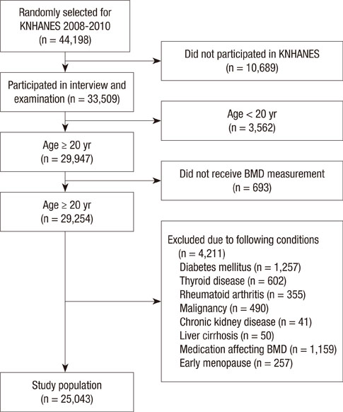
Fig. 1 Flow diagram for the identification of study population. KNHANES, Korea National Health and Nutrition Examination Survey; BMD, bone mineral density.

Fig. 2 Comparison of the prevalence of osteoporosis at various measurement sites based on the findings of this study (Korean) and established data supplied by the manufacturer of the DXA device (Hologic) in men (A) and postmenopausal women (B) ≥ 50 yr of age. Korean indicates the prevalence of osteoporosis as determined using the reference values from this study, and Hologic indicates the prevalence of osteoporosis as determined using the reference values provided by the manufacturer of the DXA device (based on a study of native Japanese subjects). DXA, dual-energy X-ray absorptiometry.
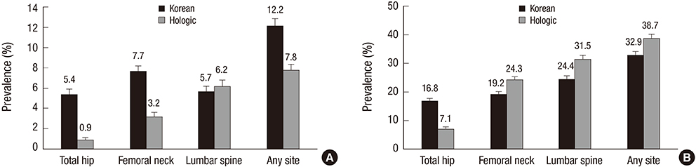
Fig. 3 Comparison of the prevalence of osteoporosis in men (A) and postmenopausal women (B) ≥ 50 yr of age in various age groups according to the different reference values. Osteoporosis was defined based on the WHO classification (T-score ≤ -2.5 standard deviation). Korean indicates the prevalence of osteoporosis as determined using the reference values from this study, and Hologic indicates the prevalence of osteoporosis as determined using the reference values provided by the manufacturer of the DXA device (based on a study of native Japanese subjects). DXA, dual-energy X-ray absorptiometry; WHO, World Health Organization.
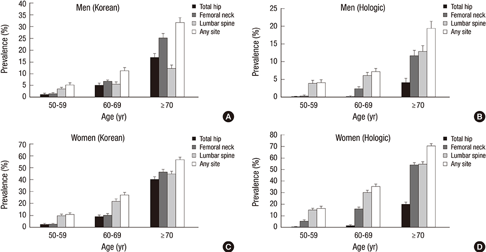
Fig. 4 Comparison of the prevalence of osteoporosis in men (A and B) and postmenopausal women (C and D) ≥ 50 yr of age in various age groups and at various measurement sites according to the different reference values. Korean indicates the prevalence of osteoporosis as determined using the reference values from this study, and Hologic indicates the prevalence of osteoporosis as determined using the reference values provided by the manufacturer of the DXA device (based on a study of native Japanese subjects). DXA, dual-energy X-ray absorptiometry.
Reference
-
1. Melton LJ 3rd. Who has osteoporosis? A conflict between clinical and public health perspectives. J Bone Miner Res. 2000; 15:2309–2314.2. Mithal A, Dhingra V, Lau E. The Asian audit: epidemiology, costs and burden of osteoporosis in Asia 2009. accessed on 2 March 2014. Available at http://www.iofbonehealth.org/asian-audit.3. Assessment of fracture risk and its application to screening for postmenopausal osteoporosis. Report of a WHO Study Group. World Health Organ Tech Rep Ser. 1994; 843:1–129.4. Looker AC, Melton LJ 3rd, Borrud LG, Shepherd JA. Lumbar spine bone mineral density in US adults: demographic patterns and relationship with femur neck skeletal status. Osteoporos Int. 2012; 23:1351–1360.5. Wu XP, Liao EY, Huang G, Dai RC, Zhang H. A comparison study of the reference curves of bone mineral density at different skeletal sites in native Chinese, Japanese, and American Caucasian women. Calcif Tissue Int. 2003; 73:122–132.6. Ho-Pham LT, Nguyen UD, Pham HN, Nguyen ND, Nguyen TV. Reference ranges for bone mineral density and prevalence of osteoporosis in Vietnamese men and women. BMC Musculoskelet Disord. 2011; 12:182.7. Cheng XG, Yang DZ, Zhou Q, Zhuo TJ, Zhang HC, Xiang J, Wang HF, Ou PZ, Liu JL, Xu L, et al. Age-related bone mineral density, bone loss rate, prevalence of osteoporosis, and reference database of women at multiple centers in China. J Clin Densitom. 2007; 10:276–284.8. Kin K, Kushida K, Yamazaki K, Okamoto S, Inoue T. Bone mineral density of the spine in normal Japanese subjects using dual-energy X-ray absorptiometry: effect of obesity and menopausal status. Calcif Tissue Int. 1991; 49:101–106.9. Yang SO, Lee SK. Normative bone mineral density data by digital X-ray radiogrammetry in Korean men. J Korean Soc Osteoporos. 2009; 7:22–27.10. Orimo H, Sugioka Y, Fukunaga M, Muto Y, Hotokebuchi T, Gorai I, Nakamura T, Kushida K, Tanaka H, Ikai T, et al. Diagnostic criteria of primary osteoporosis. J Bone Miner Metab. 1998; 16:139–150.11. Simonelli C, Adler RA, Blake GM, Caudill JP, Khan A, Leib E, Maricic M, Prior JC, Eis SR, Rosen C, et al. Dual-energy X-ray absorptiometry technical issues: the 2007 ISCD official positions. J Clin Densitom. 2008; 11:109–122.12. Wolter KM. Introduction to variance estimation. New York: Springer;1985.13. Ribom EL, Ljunggren O, Mallmin H. Use of a Swedish T-score reference population for women causes a two-fold increase in the amount of postmenopausal Swedish patients that fulfill the WHO criteria for osteoporosis. J Clin Densitom. 2008; 11:404–411.14. Mazess RB, Barden H. Bone density of the spine and femur in adult white females. Calcif Tissue Int. 1999; 65:91–99.15. Zhang ZQ, Ho SC, Chen ZQ, Zhang CX, Chen YM. Reference values of bone mineral density and prevalence of osteoporosis in Chinese adults. Osteoporos Int. 2014; 25:497–507.16. Marwaha RK, Tandon N, Kaur P, Sastry A, Bhadra K, Narang A, Arora S, Mani K. Establishment of age-specified bone mineral density reference range for Indian females using dual-energy X-ray absorptiometry. J Clin Densitom. 2012; 15:241–249.17. Lee J, Lee S, Jang S, Ryu OH. Age-related changes in the prevalence of osteoporosis according to gender and skeletal site: the Korea National Health and Nutrition Examination Survey 2008-2010. Endocrinol Metab (Seoul). 2013; 28:180–191.18. Iki M, Kagamimori S, Kagawa Y, Matsuzaki T, Yoneshima H, Marumo F. Bone mineral density of the spine, hip and distal forearm in representative samples of the Japanese female population: Japanese Population-Based Osteoporosis (JPOS) Study. Osteoporos Int. 2001; 12:529–537.19. Kudlacek S, Schneider B, Peterlik M, Leb G, Klaushofer K, Weber K, Woloszczuk W, Willvonseder R. Normative data of bone mineral density in an unselected adult Austrian population. Eur J Clin Invest. 2003; 33:332–339.20. Simmons A, Barrington S, O'Doherty MJ, Coakley AJ. Dual energy X-ray absorptiometry normal reference range use within the UK and the effect of different normal ranges on the assessment of bone density. Br J Radiol. 1995; 68:903–909.21. Lau EM, Lynn H, Woo J, Melton LJ 3rd. Areal and volumetric bone density in Hong Kong Chinese: a comparison with Caucasians living in the United States. Osteoporos Int. 2003; 14:583–588.22. Go SW, Cha YH, Lee JA, Park HS. Association between sarcopenia, bone density, and health-related quality of life in Korean men. Korean J Fam Med. 2013; 34:281–288.23. Lee DR, Lee J, Rota M, Lee J, Ahn HS, Park SM, Shin D. Coffee consumption and risk of fractures: a systematic review and dose-response meta-analysis. Bone. 2014; 63:20–28.24. Kim N, Choi HR, Kim SW, Kim BS, Won CW, Kim SY. Association between bone mineral density and sleep duration in the Korean elderly population. Korean J Fam Med. 2014; 35:90–97.25. Henry MJ, Pasco JA, Korn S, Gibson JE, Kotowicz MA, Nicholson GC. Bone mineral density reference ranges for Australian men: Geelong Osteoporosis Study. Osteoporos Int. 2010; 21:909–917.26. Looker AC, Wahner HW, Dunn WL, Calvo MS, Harris TB, Heyse SP, Johnston CC Jr, Lindsay RL. Proximal femur bone mineral levels of US adults. Osteoporos Int. 1995; 5:389–409.27. Melamed A, Vittinghoff E, Sriram U, Schwartz AV, Kanaya AM. BMD reference standards among south asians in the United States. J Clin Densitom. 2010; 13:379–384.28. Ahmed AI, Blake GM, Rymer JM, Fogelman I. Screening for osteopenia and osteoporosis: do the accepted normal ranges lead to overdiagnosis? Osteoporos Int. 1997; 7:432–438.29. Delezé M, Cons-Molina F, Villa AR, Morales-Torres J, Gonzalez-Gonzalez JG, Calva JJ, Murillo A, Briceño A, Orozco J, Morales-Franco G, et al. Geographic differences in bone mineral density of Mexican women. Osteoporos Int. 2000; 11:562–569.30. Wu XP, Liao EY, Zhang H, Dai RC, Shan PF, Cao XZ, Liu SP, Jiang Y. Determination of age-specific bone mineral density and comparison of diagnosis and prevalence of primary osteoporosis in Chinese women based on both Chinese and World Health Organization criteria. J Bone Miner Metab. 2004; 22:382–391.31. Choi SH, Park IJ, Joo NS, Kim BT. Reference value for the T-score in osteoporosis diagnosis by health screening subjects. Korean J Bone Metab. 2008; 15:67–76.32. Kanis JA, Glüer CC. An update on the diagnosis and assessment of osteoporosis with densitometry. Committee of Scientific Advisors, International Osteoporosis Foundation. Osteoporos Int. 2000; 11:192–202.33. Cui LH, Choi JS, Shin MH, Kweon SS, Park KS, Lee YH, Nam HS, Jeong SK, Im JS. Prevalence of osteoporosis and reference data for lumbar spine and hip bone mineral density in a Korean population. J Bone Miner Metab. 2008; 26:609–617.34. Kanis JA, Johnell O, Oden A, Jonsson B, De Laet C, Dawson A. Risk of hip fracture according to the World Health Organization criteria for osteopenia and osteoporosis. Bone. 2000; 27:585–590.35. Kanis JA, McCloskey EV, Johansson H, Oden A, Melton LJ 3rd, Khaltaev N. A reference standard for the description of osteoporosis. Bone. 2008; 42:467–475.36. Yoon HK, Park C, Jang S, Jang S, Lee YK, Ha YC. Incidence and mortality following hip fracture in Korea. J Korean Med Sci. 2011; 26:1087–1092.37. Khosla S. Update in male osteoporosis. J Clin Endocrinol Metab. 2010; 95:3–10.38. Abrahamsen B. Adverse effects of bisphosphonates. Calcif Tissue Int. 2010; 86:421–435.39. Henzell S, Dhaliwal SS, Price RI, Gill F, Ventouras C, Green C, Da Fonseca F, Holzherr M, Prince R. Comparison of pencil-beam and fan-beam DXA systems. J Clin Densitom. 2003; 6:205–210.
- Full Text Links
-
- Actions
-
Cited
- CITED
-
- Close
- Share
-
- Similar articles
-
- Prevalence of Osteoporosis in the Korean Population Based on Korea National Health and Nutrition Examination Survey (KNHANES), 2008-2011
- Factors associated with the bone mineral density in Korean adults: Data from the 2010-2011 Korean National Health and Nutrition Examination Survey (KNHANES) V
- Milk Consumption and Bone Mineral Density in Adults: Using Data from the Korea National Health and Nutrition Examination Survey 2008–2011
- Association between Coffee Consumption and Bone Mineral Density in Korean Men Aged 50 Years and Older: A Cross Sectional Analysis of Korea National Health and Nutrition Examination Survey 2011
- Vitamin D intake and bone mineral density in Korean adults: analysis of the 2009–2011 Korea National Health and Nutrition Examination Survey
