Pediatr Gastroenterol Hepatol Nutr.
2015 Sep;18(3):193-196. 10.5223/pghn.2015.18.3.193.
Ileal Fecaloma Presenting with Small Bowel Obstruction
- Affiliations
-
- 1Department of Pediatrics, Konkuk University Medical Center, Konkuk University School of Medicine, Seoul, Korea. baedori@hanafos.com
- 2Department of Surgery, Konkuk University Medical Center, Konkuk University School of Medicine, Seoul, Korea.
- KMID: 2068797
- DOI: http://doi.org/10.5223/pghn.2015.18.3.193
Abstract
- A fecaloma refers to a mass of accumulated feces that is much harder than a mass associated with fecal impaction. Fecalomas are usually found in the rectosigmoid area. A 10-year-old male with chronic constipation was admitted because of increasing abdominal pain. An abdominal computed tomography scan and a simple abdominal x-ray revealed rapidly evolving mechanical obstruction in the small intestine. Most of the fecalomas are successfully treated by conservative methods such as laxatives, enemas and rectal evacuation. When conservative treatments have failed, surgical intervention may be needed. In this case, an emergency operation was performed and a 4x3x2.5 cm fecaloma was found in the distal ileum. We thus report a case of ileal fecaloma inducing small bowel obstruction in a patient with chronic constipation, who required surgical intervention. When symptoms of acute small intestinal obstruction develop in a patient with chronic constipation, a fecaloma should be considered in differential diagnosis.
Keyword
MeSH Terms
Figure
-
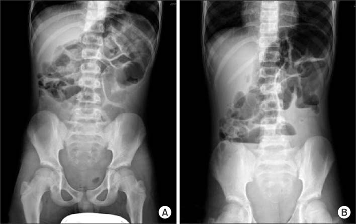
Fig. 1 A plain x-ray of the abdomen (A: supine view, B: erect view) showing a mechanical small bowel obstruction with 'step-ladder' air-fluid pattern in the erect film.
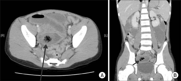
Fig. 2 Abdominopelvic computed tomography scan (A: axial view, B: coronal view) showing the 1.8 cm-sized round intraluminal hypodense mass (black arrows) with a 'mottled gas' pattern in the lumen of distal ileum.

Fig. 3 The fecaloma. A 4×3×2.5 cm-sized fecaloma was found in the distal ileum at 65 cm proximal to the ileocecal valve, and removed through enterotomy.
Reference
-
1. Garisto JD, Campillo L, Edwards E, Harbour M, Ermocilla R. Giant fecaloma in a 12-year-old-boy: a case report. Cases J. 2009; 2:127.
Article2. Cid AA, Pietruk T, Bidari CZ, Ehrinpreis MN. Cecal fecaloma mimicking colonic neoplasm. Dig Dis Sci. 1981; 26:1134–1137.
Article3. Gilbert RF. Cecal infarction secondary to a distal obstructing fecaloma: association with drug abuse. South Med J. 1980; 73:1296–1297.4. Sakai E, Inokuchi Y, Inamori M, Uchiyama T, Iida H, Takahashi H, et al. Rectal fecaloma: successful treatment using endoscopic removal. Digestion. 2007; 75:198.
Article5. Park JS, Park TJ, Hwa JS, Seo JH, Park CH, Youn HS. Acute urinary retention in a 47-month-old girl caused by the giant fecaloma. Pediatr Gastroenterol Hepatol Nutr. 2013; 16:200–205.
Article6. Yucel AF, Akdogan RA, Gucer H. A giant abdominal mass: fecaloma. Clin Gastroenterol Hepatol. 2012; 10:e9–e10.
Article7. Sonnenberg A, Koch TR. Physician visits in the United States for constipation: 1958 to 1986. Dig Dis Sci. 1989; 34:606–611.
Article8. Kim SM, Ryu KH, Kim YS, Lee TH, Im EH, Huh KC, et al. Cecal fecaloma due to intestinal tuberculosis: endoscopic treatment. Clin Endosc. 2012; 45:174–176.
Article9. Rajagopal A, Martin J. Giant fecaloma with idiopathic sigmoid megacolon: report of a case and review of the literature. Dis Colon Rectum. 2002; 45:833–835.
- Full Text Links
-
- Actions
-
Cited
- CITED
-
- Close
- Share
-
- Similar articles
-
- Primary Malignant Fibrous Histiocytoma (MFH) of the Small Bowel Presenting as an Intussusception Causing Small Bowel Obstruction
- Congenital Ileal Atresia Presenting as a Single Cyst-like Dilated Bowel on Prenatal Sonography at Late Third Trimester: A Case Report
- Small bowel intubation using guide wire: use in decompression of small bowel obstruction
- A Transmesenteric Hernia in a Child: Gangrene of a Long Segment of Small Bowel through a Large Mesenteric Defect
- A Case of Recurrent Intestinal Obstruction Caused by Meckel's Diverticulum
