J Korean Soc Radiol.
2015 Oct;73(4):259-263. 10.3348/jksr.2015.73.4.259.
Spontaneous Infarction of Benign Breast Lesion during Pregnancy: Ultrasonographic and Pathologic Findings
- Affiliations
-
- 1Department of Radiology, Eulji University Hospital, Daejeon, Korea. kskim@eulji.ac.kr
- 2Department of Radiology, Health Care Center, Pohang, Korea.
- 3Department of Pathology, Eulji University Hospital, Daejeon, Korea.
- KMID: 2068717
- DOI: http://doi.org/10.3348/jksr.2015.73.4.259
Abstract
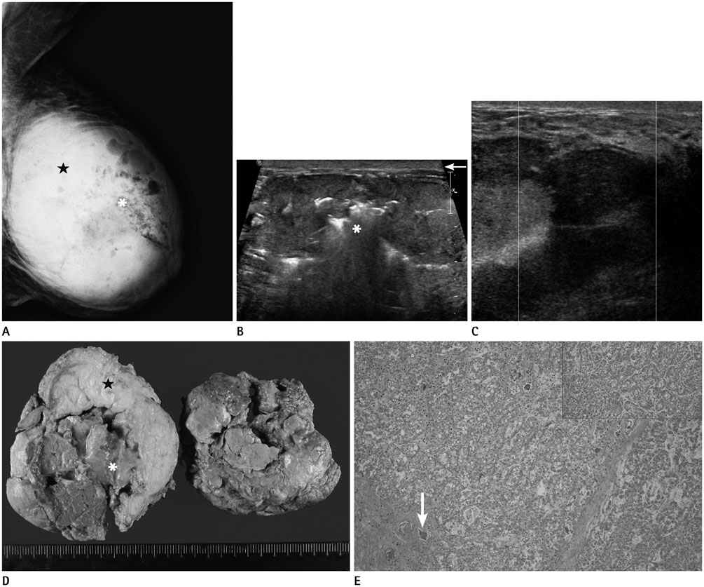
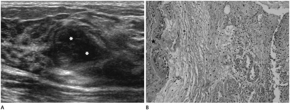
- The spontaneous infarction of benign breast lesions is a rare entity and hence is not usually considered in the differential diagnosis during radiologic or clinical examination. There have been a few published cases of infarction during pregnancy and lactation. In this study we report the ultrasonographic and pathologic features of a spontaneous infarction of a lactating adenoma with acute mastitis and abscess and a spontaneously infarcted fibroadenoma.
MeSH Terms
Figure
Reference
-
1. Behrndt VS, Barbakoff D, Askin FB, Brem RF. Infarcted lactating adenoma presenting as a rapidly enlarging breast mass. AJR Am J Roentgenol. 1999; 173:933–935.2. Sabate JM, Clotet M, Torrubia S, Gomez A, Guerrero R, de las Heras P, et al. Radiologic evaluation of breast disorders related to pregnancy and lactation. Radiographics. 2007; 27:Suppl 1. S101–S124.3. Skenderi F, Krakonja F, Vranic S. Infarcted fibroadenoma of the breast: report of two new cases with review of the literature. Diagn Pathol. 2013; 8:38.4. Oh YJ, Choi SH, Chung SY, Yang I, Woo JY, Lee MJ. Spontaneously infarcted fibroadenoma mimicking breast cancer. J Ultrasound Med. 2009; 28:1421–1423.5. Lucey JJ. Spontaneous infarction of the breast. J Clin Pathol. 1975; 28:937–943.6. Oh HJ, Kim SH, Kang BJ, Lee AW, Song BJ, Kim HS, et al. Ultrasonographic features of spontaneous breast tumor infarction. Breast Cancer. 2014; Mar. 15. [Epub]. DOI: 10.1007/s12282-014-0525-3.7. Kim SJ. Spontaneously infarcted fibroadenoma of the breast in an adolescent girl: sonographic findings. J Med Ultrasonics. 2014; 41:83–85.8. Hsu S, Hsieh H, Hsu G, Lee H, Chen K, Yu J. Spontaneous infarction of a fibroadenoma of the breast in a 12-year-old girl. J Med Sci Taipei. 2005; 25:313.9. Chang YW, Kwon KH, Goo DE, Choi DL, Lee HK, Yang SB. Sonographic differentiation of benign and malignant cystic lesions of the breast. J Ultrasound Med. 2007; 26:47–53.10. Fowler CL. Spontaneous infarction of fibroadenoma in an adolescent girl. Pediatr Radiol. 2004; 34:988–990.
- Full Text Links
- Actions
-
Cited
- CITED
-
- Close
- Share
- Similar articles
-
- Ultrasonographic Findings of Breast Diseases During Pregnancy and Lactating Period
- Spontaneous Infarction of Hyperplastic Breast Tissue: A Case Report
- Spontaneous Infarction of Phyllodes Tumor of the Breast in a Postpartum Woman: A Case Report
- Pregnancy-Associated Breast Disease: Radiologic Features and Diagnostic Dilemmas
- The analysis of ultrasonographic findings in breast carcinoma


