Spontaneous Infarction of Phyllodes Tumor of the Breast in a Postpartum Woman: A Case Report
- Affiliations
-
- 1Department of Radiology, Wonkwang University School of Medicine, Wonkwang University Hospital, Iksan, Korea. khw@wonkwang.ac.kr
- 2Department of Pathology, Wonkwang University School of Medicine, Wonkwang University Hospital, Iksan, Korea.
- 3Department of Surgery, Wonkwang University School of Medicine, Wonkwang University Hospital, Iksan, Korea.
- KMID: 2394047
- DOI: http://doi.org/10.3348/jksr.2017.77.5.327
Abstract
- A rare tumor of the breast, phyllodes tumor is uncommon in pregnant women, and spontaneous infarction of this tumor has not been reported to date. Infarction develops in malignant tumors of the breast, but the mechanism and pathogenesis of this complication is not fully understood. Breast tumor infarction in pregnant women is uncommon, except in cases of fibroadenomas. The authors report a case of spontaneous infarction of a benign phyllodes tumor in a 30-year-old postpartum woman that exhibited rapid growth during late pregnancy; this is followed by a discussion of imaging findings.
MeSH Terms
Figure
-
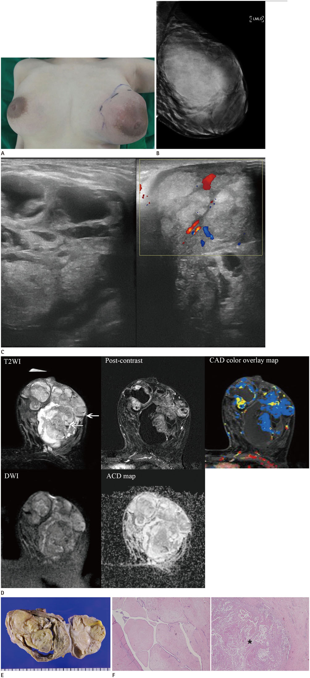
Fig. 1 A 30-year-old postpartum woman with spontaneous infarction of phylloides tumor, presenting as a huge palpable non-tender mass in her left breast. A. Clinical photograph shows a hard, palpable mass with erythematous skin change in a bulging left breast. B. Mammography shows a huge oval hyperdense mass in the left breast. C. US reveals a large circumscribed mass with a multiloculated cystic portion, and color doppler US shows penetrating vascularity within the solid portion of the mass. US = ultrasound D. Magnetic resonance imaging of the left breast mass: T2WI shows a multiloculated heterogeneously iso- to high signal intensity mass with dark septations, containing a cystic portion and scant hemorrhagic fluid collection (arrows). Fat suppressed subtraction image at 1-minute post-gadolinium injection shows heterogeneous enhancement and a smooth wall of the mass. CAD color overlay map obtained by dynamic contrast enhancement study shows that the most of the solid portion displayed a persistent kinetic pattern. DWI and corresponding ADC map show no diffusion restriction or hyperintensity. E. Gross photograph shows a 14 × 11.5 × 8 cm in size and yellow colored mass. A section of the mass revealed a multicystic lesion with inflammation and necrosis. F. Microscopic examination shows a tumor composed of mesenchymal nodules and leaf-like projections with intervening clefts. Hematoxylin and eosin staining (× 40) shows coagulation necrosis (asterisks) in the mass induced by infarction. ADC = apparent diffusion coefficient, CAD = computer aided detection, DWI = diffusion-weighted image, T2WI = T2-weighted image
Reference
-
1. Murthy SS, Raju KV, Nair HG. Phyllodes tumor in a lactating breast. Clin Med Insights Pathol. 2016; 9:13–17.2. Behrndt VS, Barbakoff D, Askin FB, Brem RF. Infarcted lactating adenoma presenting as a rapidly enlarging breast mass. AJR Am J Roentgenol. 1999; 173:933–935.3. Kim JY, Kim KS, Lee Y, Kim JH. Spontaneous Infarction of benign breast lesion during pregnancy: ultrasonographic and pathologic findings. J Korean Soc Radiol. 2015; 73:259–263.4. Likhitmaskul T, Asanprakit W, Charoenthammaraksa S, Lohsiriwat V, Supaporn S, Vassanasiri W, et al. Giant benign phyllodes tumor with lactating changes in pregnancy: a case report. Gland Surg. 2015; 4:339–343.5. Kim SJ. Spontaneously infarcted fibroadenoma of the breast in an adolescent girl: sonographic findings. J Med Ultrason (2001). 2014; 41:83–85.6. Jesinger RA, Lattin GE Jr, Ballard EA, Zelasko SM, Glassman LM. Vascular abnormalities of the breast: arterial and venous disorders, vascular masses, and mimic lesions with radiologic-pathologic correlation. Radiographics. 2011; 31:E117–E136.7. Oh HJ, Kim SH, Kang BJ, Lee AW, Song BJ, Kim HS, et al. Ultrasonographic features of spontaneous breast tumor infarction. Breast Cancer. 2015; 22:596–601.8. Verslegers I, Tjalma W, Van Goethem M, Colpaert C, Biltjes I, De Schepper AM, et al. Massive infarction of a recurrent phyllodes tumor of the breast: MRI-findings. JBR-BTR. 2004; 87:21–22.9. Yabuuchi H, Soeda H, Matsuo Y, Okafuji T, Eguchi T, Sakai S, et al. Phyllodes tumor of the breast: correlation between MR findings and histologic grade. Radiology. 2006; 241:702–709.10. Tan H, Zhang S, Liu H, Peng W, Li R, Gu Y, et al. Imaging findings in phyllodes tumors of the breast. Eur J Radiol. 2012; 81:e62–e69.
- Full Text Links
-
- Actions
-
Cited
- CITED
-
- Close
- Share
-
- Similar articles
-
- Myxoid Liposarcoma of the Breast Mimicking Phyllodes Tumor: A Case Report
- A case of phyllodes tumor in a 10-year-old girl
- Ductal Carcinoma In Situ Arising in a Benign Phyllodes Tumor: A Case Report
- Spontaneous Infarction of Hyperplastic Breast Tissue: A Case Report
- Invasive Cribriform Carcinoma Arising in Malignant Phyllodes Tumor of Breast: A Case Report
