J Korean Soc Radiol.
2015 Sep;73(3):168-171. 10.3348/jksr.2015.73.3.168.
Spontaneous Infarction of Hyperplastic Breast Tissue: A Case Report
- Affiliations
-
- 1Department of Radiology, Gil Medical Center, Gachon University School of Medicine and Science, Incheon, Korea. sangyu.nam7@gmail.com
- KMID: 2043607
- DOI: http://doi.org/10.3348/jksr.2015.73.3.168
Abstract
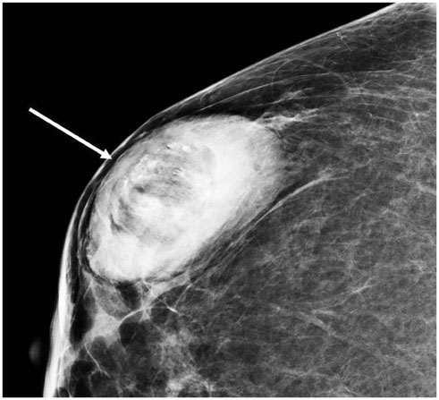
- Spontaneous breast infarction is a very rare complication of fibroadenoma of the breast. We present an interesting case of a 33-year-old woman with spontaneous infarction of hyperplastic breast tissue related to pregnancy and lactation. Mammography showed an oval, circumscribed, fat-containing mass with microcalcifications. Ultrasonography revealed an oval, circumscribed mass with echogenic dots. Color Doppler imaging revealed presence of minimal vascularity at the periphery of the mass.
MeSH Terms
Figure
Reference
-
1. Oh HJ, Kim SH, Kang BJ, Lee AW, Song BJ, Kim HS, et al. Ultrasonographic features of spontaneous breast tumor infarction. Breast Cancer. 2014; 03. 15. DOI: 10.1007/s12282-014-0525-3.2. Lucey JJ. Spontaneous infarction of the breast. J Clin Pathol. 1975; 28:937–943.3. Raju GC, Naraynsingh V. Infarction of fibroadenoma of the breast. J R Coll Surg Edinb. 1985; 30:162–163.4. Skenderi F, Krakonja F, Vranic S. Infarcted fibroadenoma of the breast: report of two new cases with review of the literature. Diagn Pathol. 2013; 8:38.5. Behrndt VS, Barbakoff D, Askin FB, Brem RF. Infarcted lactating adenoma presenting as a rapidly enlarging breast mass. AJR Am J Roentgenol. 1999; 173:933–935.6. Jimenez JF, Ryals RO, Cohen C. Spontaneous breast infarction associated with pregnancy presenting as a palpable mass. J Surg Oncol. 1986; 32:174–178.7. Fowler CL. Spontaneous infarction of fibroadenoma in an adolescent girl. Pediatr Radiol. 2004; 34:988–990.8. Deshpande KM, Deshpande AH, Raut WK, Lele VR, Bobhate SK. Diagnostic difficulties in spontaneous infarction of a fibroadenoma in an adolescent: case report. Diagn Cytopathol. 2002; 26:26–28.9. Greenberg ML, Middleton PD, Bilous AM. Infarcted intraduct papilloma diagnosed by fine-needle biopsy: a cytologic, clinical, and mammographic pitfall. Diagn Cytopathol. 1994; 11:188–191. discussion 191-19410. Pinto RG, Couto F, Mandreker S. Infarction after fine needle aspiration. A report of four cases. Acta Cytol. 1996; 40:739–741.
- Full Text Links
- Actions
-
Cited
- CITED
-
- Close
- Share
- Similar articles
-
- Spontaneous Infarction of Phyllodes Tumor of the Breast in a Postpartum Woman: A Case Report
- Spontaneous Infarction of Benign Breast Lesion during Pregnancy: Ultrasonographic and Pathologic Findings
- Spontaneous Regression of Hyperplastic Gastric Polyps
- Unilateral autoinflation of a saline-filled breast implant initially diagnosed as capsular contracture: a case report and review of the literature
- A Case of Gastric Hyperplastic Polyposis Associated with Colonic Hyperplastic Polyposis


