Pregnancy-Associated Breast Disease: Radiologic Features and Diagnostic Dilemmas
- Affiliations
-
- 1Department of Diagnostic Radiology, The Research Institute of Radiological Science, Yonsei University College of Medicine, Seoul, Korea. ekkim@yumc.yonsei.ac.kr
- KMID: 1715871
- DOI: http://doi.org/10.3349/ymj.2006.47.1.34
Abstract
- In this paper, we evaluate the radiological features of pregnancy-associated breast lesions and discuss the difficulties in diagnosis by imaging. We selected patients who were diagnosed with pregnancy-associated breast lesions during the previous 5 years. All patients complained of palpable lesions in the breast and underwent ultrasonographic (US) examination, the first choice for examination of pregnancy-related breast lesions. Any suspicious lesions found by the US were recommended for a US-guided core biopsy, US-guided fine needle aspiration (FNA), or surgery. Various breast lesions were detected during pregnancy and lactation, including breast cancer, mastitis and abscesses, lactating adenoma, galactoceles, lobular hyperplasia, and fibroadenomas. The imaging features of pregnancy-associated breast lesions did not differ from the features of non-pregnancy-associated breast lesions; however, some pregnancy-associated benign lesions had suspicious sonographic features. A US-guided core biopsy was necessary for differentiating benign from malignant. In patients with breast cancer, the cancer was often advanced at the time of diagnosis. In conclusion, various pregnancy-related breast lesions were detected and the imaging of these lesions had variable findings. Breast ultrasound could be an excellent imaging modality for diagnosis and differentiation between benign and malignant lesions. However, when the imaging results are suspicious, a biopsy should be performed to obtain a pathologic diagnosis.
MeSH Terms
Figure
-

Fig. 1 Ultrasound of a 28-year-old lactating woman (postpartum 6 months) without breast disease. The images show an ill-defined, low echoic lesion, suggesting duct dilatation (white arrows) due to pregnancy.
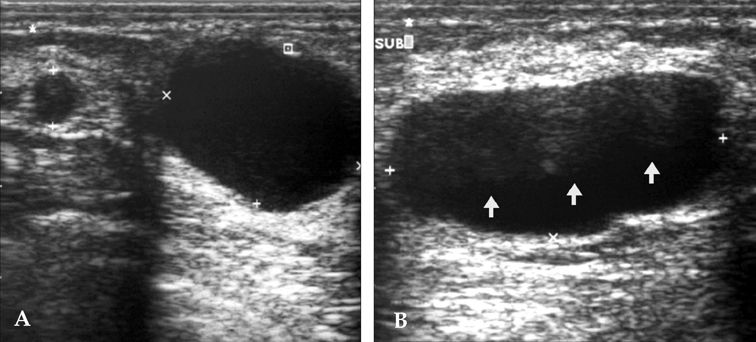
Fig. 2 Ultrasound of a 35-year-old lactating woman (postpartum 6 months) with galactoceles. (A) There are two anechoic, cyst-like masses. The larger mass shows posterior acoustic enhancement with lateral edge shadowing, suggesting a pure cystic galactocele by clinical history. (B) Another cystic mass shows a fat-fluid level (arrows) representing a galactocele.
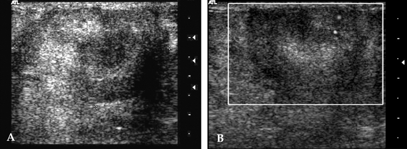
Fig. 3 Images from a 35-year-old lactating woman (postpartum 4 months) with a galactocele. (A) US of the breast shows a lobulated and heterogeneous, echoic mass in the palpable area. (B) The Doppler study shows increased blood flow in the mass. This lesion was confirmed to be a galactocele by US-guided core biopsy.

Fig. 4 US of a 38-year-old lactating woman (postpartum 6 months) with a galactocele. US of the breast reveal an irregular-shaped, hypoechoic mass in the left subareolar area (white arrows). Part of the mass shows spiculation. Under the diagnosis of category 4A, a core biopsy was performed, and the pathology result was fibrocystic changes, compatible with a galactocele.
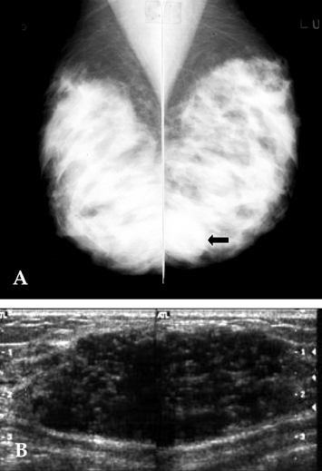
Fig. 5 Images of a 30-year-old woman (postpartum 5 months) with a lactating adenoma. (A) Both oblique mammograms show bilateral, extremely dense breast patterns with a well-circumscribed, high-density mass in the low-posterior portion of the left breast (arrow). (B) US reveals a large, relatively well-defined, oval-shaped, inhomogeneous, hypoechoic mass.

Fig. 6 US of a 29-year-old woman (postpartum 4 months) with a lactating adenoma. US shows a 2-cm, ill-defined, hypoechoic lesion which was categorized as a suspicious lesion (category 4A) and followed-up with a US-guided core biopsy. The histopathological diagnosis was a lactating adenoma.
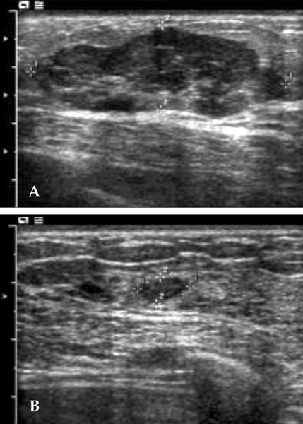
Fig. 7 Images from a 28-year-old pregnant woman (IUP 36 weeks) with a lactating adenoma. (A) US shows an approximately 4.5 × 1.5-cm, lobular-shaped, well-circumscribed, inhomogeneous, hypoechoic mass in the left breast. A US-guided biopsy was performed which confirmed the lesion was a lactating adenoma. (B) The follow-up sonography was taken 6 months later, after delivery. The lactating adenoma had decreased in size to less than 1 cm.
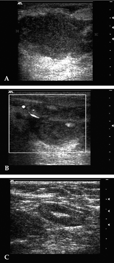
Fig. 8 Images from a 29-year-old woman (postpartum 2 weeks) with breast abscess. (A) US of the left breast shows an irregular-shaped, heterogeneous, hypoechoic mass-like lesion with indistinct margins and partial posterior enhancement at the subareolar portion. Internal moving debris was also noted, and approximately 20 cc of pus was drained through an 18-G needle. (B) On Doppler, marked increased blood flow is seen at the margin of the abscess. (C) A cortical thickened, enlarged lymph node is noted in the left axilla.
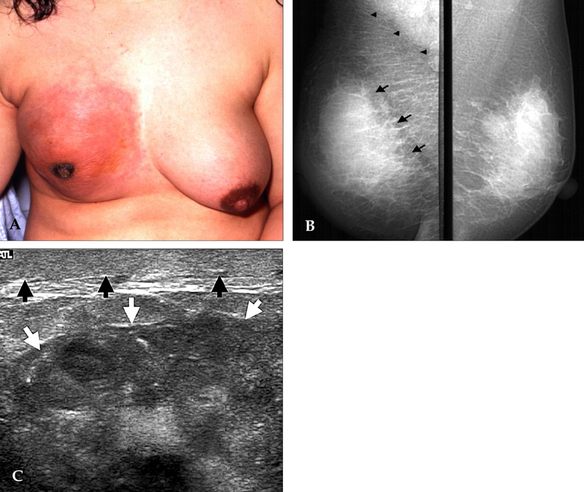
Fig. 9 Images from a 33-year-old woman (postpartum 3 weeks) with advanced breast carcinoma. The patient noticed painful swelling of the right breast during the 20th week of pregnancy, but felt that these were normal physiological changes. (A) The right breast shows a discolored and contracted appearance with nipple retraction, compared with the left breast. (B) Mammography shows a huge mass in the entire right breast (arrows), with diffuse trabecular thickening by lymphatic engorgement and multiple metastatic lymph nodes in the axilla (arrow heads). (C) US shows an irregular-shaped, inhomogeneous, echoic mass (white arrows) with diffuse skin thickening (black arrows).
Cited by 1 articles
-
Breast diseases during pregnancy and lactation
Ji Hoon Yu, Min Jeong Kim, Hyonil Cho, Hyun Ju Liu, Sei-Jun Han, Tae-Gyu Ahn
Obstet Gynecol Sci. 2013;56(3):143-159. doi: 10.5468/ogs.2013.56.3.143.
Reference
-
1. Smith MS. Patton HD, Fuchs AF, Mille B, Scher AM, Steiner R, editors. Lactation. Textbook of physiology. 1989. 21st ed. Philadelphia: Saunders;1408–1421.2. Kopans DB. Breast imaging. 1998. 2th ed. Philadelphia: Lippincott-Raven;445–496.3. Golden GT, Wangensteen SL. Galactocele of the breast. Am J Surg. 1972. 123:271–273.4. Sawhney S, Petkovska L, Ramadan S, Al-Muhtaseb S, Jain R, Sheikh M. Sonographic appearances of galactoceles. J Clin Ultrasound. 2002. 30:18–22.5. Park MS, Oh KK, Kim EK, Lee SI. Multifaces of sonographic findings of galactocele: comparison according to its association with pregnancy. J Korean Radiol Soc. 2000. 42:699–703.6. Slavin JL, Billson VR, Ostor AG. Nodular breast lesions during pregnancy and lactation. Histopathology. 1993. 22:481–485.7. Sumkin JH, Perrone AM, Harris KM, Nath ME, Amortegui AJ, Weinstein BJ. Lactating adenoma: US features and literature review. Radiology. 1998. 206:271–274.8. Darling ML, Smith DN, Rhei E, Denison CM, Lester SC, Meyer JE. Lactating adenoma: sonographic features. Breast J. 2000. 6:252–256.9. Kaufmann R, Foxman B. Mastitis among lactating women: occurrence and risk factors. Soc Sci Med. 1991. 33:701–705.10. Petrek JA. Breast cancer during pregnancy. Cancer. 1994. 74:518–527.11. Ahn BY, Kim HH, Moon WK, Pisano ED, Kim HS, Cha ES, et al. Pregnancy-and lactation-associated breast cancer: mammographic and sonographic findings. J Ultrasound Med. 2003. 22:491–499.12. Gallenberg MM, Loprinzi CL. Breast cancer and pregnancy. Semin Oncol. 1989. 16:369–376.13. Mendelson EB, Berg WA, Merritt CRB. Miller WT, Berg WA, editors. Toward a standardized breast ultrasound lexicon, BI-RADS: ultrasound. Seminars in roentgenology: Breast imaging. 2001. Vol 36. Philadelphia, PA: WB Saunders Co;217–225.14. James K, Bridger J, Anthony PP. Breast tumour of pregnancy ('lactating adenoma'). J Pathol. 1988. 156:37–44.15. Devereux WP. Acute puerperal mastitis. Evaluation of its management. Am J Obstet Gynecol. 1970. 108:78–81.16. Hook GW, Ikeda DM. Treatment of breast abscesses with US-guided percutaneous needle drainage without indwelling catheter placement. Radiology. 1999. 213:579–582.17. DiFronzo LA, O'Connell TX. Breast cancer in pregnancy and lactation. Surg Clin North Am. 1996. 76:267–278.18. Liberman L, Giess CS, Dershaw DD, Deutch BM, Petrek JA. Imaging of pregnancy-associated breast cancer. Radiology. 1994. 191:245–248.
- Full Text Links
-
- Actions
-
Cited
- CITED
-
- Close
- Share
-
- Similar articles
-
- Breast Cancer During Pregnancy
- Breast Cancer during Pregnancy
- Ultrasonographic Findings of Apocrine Lesions Arising from the Breast
- Spontaneous Infarction of Benign Breast Lesion during Pregnancy: Ultrasonographic and Pathologic Findings
- Radiologic Diagnosis of Nontuberculous Mycobacterial Pulmonary Disease
