Characterization of mandibular molar root and canal morphology using cone beam computed tomography and its variability in Belgian and Chilean population samples
- Affiliations
-
- 1Department of Imaging and Pathology, OMFS-IMPATH Research Group, Katholieke Universiteit Leuven, Leuven, Belgium. andresedvardo.torresgarcia@uzleuven.be
- 2Department of Conservative Dentistry, Katholieke Universiteit Leuven, Leuven, Belgium.
- 3Department of Endodontics, Universidad de los Andes, Santiago, Chile.
- 4Department of Oral and Maxillofacial Radiology, Universidad de los Andes, Santiago, Chile.
- KMID: 2054211
- DOI: http://doi.org/10.5624/isd.2015.45.2.95
Abstract
- PURPOSE
This study used cone-beam computed tomography (CBCT) to characterize mandibular molar root and canal morphology and its variability in Belgian and Chilean population samples.
MATERIALS AND METHODS
We analyzed the CBCT images of 515 mandibular molars (257 from Belgium and 258 from Chile). Molars meeting the inclusion criteria were analyzed to determine (1) the number of roots; (2) the root canal configuration; (3) the presence of a curved canal in the cross-sectional image of the distal root in the mandibular first molar and (4) the presence of a C-shaped canal in the second mandibular molar. A descriptive analysis was performed. The association between national origin and the presence of a curved or C-shaped canal was evaluated using the chi-squared test.
RESULTS
The most common configurations in the mesial root of both molars were type V and type III. In the distal root, type I canal configuration was the most common. Curvature in the cross-sectional image was found in 25% of the distal canals of the mandibular first molars in the Belgian population, compared to 11% in the Chilean population. The prevalence of C-shaped canals was 10% or less in both populations.
CONCLUSION
In cases of unclear or complex root and canal morphology in the mandibular molars, CBCT imaging might assist endodontic specialists in making an accurate diagnosis and in treatment planning.
MeSH Terms
Figure
-
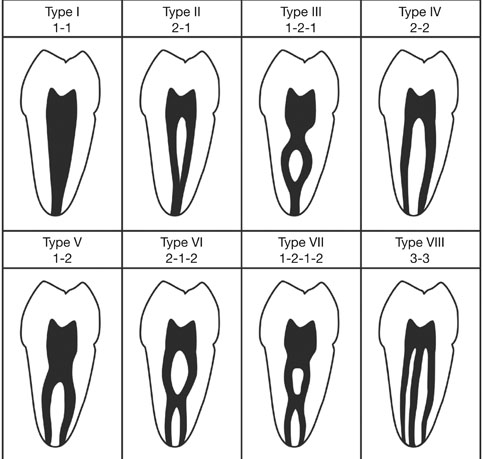
Fig. 1 Classification of root canal configuration.1 Type I: a single canal extends from the pulp chamber to the apex. Type II: two separate canals leave the pulp chamber and join short of the apex to form one canal. Type III: one canal leaves the pulp chamber, divides into two within the root, and then merges to exit as one canal. Type IV: Two separate and distinct canals extend from the pulp chamber to the apex. Type V: one canal leaves the pulp chamber and divides short of the apex into two separate and distinct canals with separate apical foramina. Type VI: two separate canals leave the pulp chamber, merge in the body of the root, and re-divide short of the apex to exit as two distinct canals. Type VII: one canal leaves the pulp chamber, divides and then rejoins within the body of the root, and finally re-divides into two distinct canals short of the apex. Type VIII: three separate and distinct canals extend from the pulp chamber to the apex.
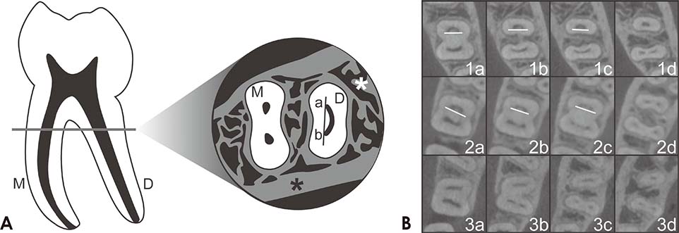
Fig. 2 Curved canal in the cross-sectional image of the distal root in the mandibular first molar. A. Left: a mandibular first molar with (M) mesial and (D) distal roots. Right: cross-sectional image of a mandibular first molar as seen in an axial-plane CBCT view. (M) mesial root, (D) distal root, (*), cortical and trabecular bone structure. When a straight line is drawn in the distal ribbon-shaped canal, it only comes in contact with the (a) buccal and (b) lingual ends of the canal. B. Cone-beam computed tomography cross-sectional images. 1a-c: curved distal canals in cross-sectional images, 1d: an oval canal, 2a and b: curved distal canals in cross-sectional images, 2c: a canal starting to divide, 2d: a fully divided canal, 3a-d: the absence of a curved distal canal in cross-sectional images.
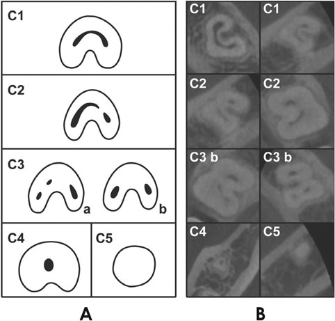
Fig. 3 Classification of C-shaped canal configurations.19 A. C1: an uninterrupted "C" with no separation or division, C2: a canal shape resembling a semi-column resulting from the discontinuation of the "C" outline, C3: three (a) or two (b) separate canals, C4: only one round or oval canal in the cross-sectional image, C5: no observable canal lumen (usually only found near the apex). B. Cross-sectional CBCT images of different types of C-shaped canal configurations, at different root levels.
Reference
-
1. Vertucci FJ. Root canal anatomy of the human permanent teeth. Oral Surg Oral Med Oral Pathol. 1984; 58:589–599.
Article2. Vertucci FJ. Root canal morphology and its relationship to endodontic procedures. Endod Topics. 2005; 10:3–29.
Article3. Mărgărit R, Andrei OC. Anatomical variations of mandibular first molar and their implications in endodontic treatment. Rom J Morphol Embryol. 2011; 52:1389–1392.4. Zaatar EI, al-Kandari AM, Alhomaidah S, al-Yasin IM. Frequency of endodontic treatment in Kuwait: radiographic evaluation of 846 endodontically treated teeth. J Endod. 1997; 23:453–456.
Article5. Scavo R, Martinez Lalis R, Zmener O, Dipietro S, Grana D, Pameijer CH. Frequency and distribution of teeth requiring endodontic therapy in an Argentine population attending a specialty clinic in endodontics. Int Dent J. 2011; 61:257–260.
Article6. Touré B, Faye B, Kane AW, Lo CM, Niang B, Boucher Y. Analysis of reasons for extraction of endodontically treated teeth: a prospective study. J Endod. 2011; 37:1512–1515.
Article7. de Pablo OV, Estevez R, Peix Sanchez M, Heilborn C, Cohenca N. Root anatomy and canal configuration of the permanent mandibular first molar: a systematic review. J Endod. 2010; 36:1919–1931.
Article8. Cotton TP, Geisler TM, Holden DT, Schwartz SA, Schindler WG. Endodontic applications of cone-beam volumetric tomography. J Endod. 2007; 33:1121–1132.
Article9. Patel S. New dimensions in endodontic imaging: part 2. Cone beam computed tomography. Int Endod J. 2009; 42:463–475.
Article10. Damstra J, Fourie Z, Huddleston Slater JJ, Ren Y. Accuracy of linear measurements from cone-beam computed tomography-derived surface models of different voxel sizes. Am J Orthod Dentofacial Orthop. 2010; 137:16.e1–16.e6.
Article11. Stratemann SA, Huang JC, Maki K, Miller AJ, Hatcher DC. Comparison of cone beam computed tomography imaging with physical measures. Dentomaxillofac Radiol. 2008; 37:80–93.
Article12. Ludlow JB, Laster WS, See M, Bailey LJ, Hershey HG. Accuracy of measurements of mandibular anatomy in cone beam computed tomography images. Oral Surg Oral Med Oral Pathol Oral Radiol Endod. 2007; 103:534–542.
Article13. Pinsky HM, Dyda S, Pinsky RW, Misch KA, Sarment DP. Accuracy of three-dimensional measurements using cone-beam CT. Dentomaxillofac Radiol. 2006; 35:410–416.
Article14. Neelakantan P, Subbarao C, Subbarao CV. Comparative evaluation of modified canal staining and clearing technique, conebeam computed tomography, peripheral quantitative computed tomography, spiral computed tomography, and plain and contrast medium-enhanced digital radiography in studying root canal morphology. J Endod. 2010; 36:1547–1551.
Article15. Wang Y, Zheng QH, Zhou XD, Tang L, Wang Q, Zheng GN, et al. Evaluation of the root and canal morphology of mandibular first permanent molars in a western Chinese population by cone-beam computed tomography. J Endod. 2010; 36:1786–1789.
Article16. Zhang R, Wang H, Tian YY, Yu X, Hu T, Dummer PM. Use of cone-beam computed tomography to evaluate root and canal morphology of mandibular molars in Chinese individuals. Int Endod J. 2011; 44:990–999.
Article17. Zheng QH, Wang Y, Zhou XD, Wang Q, Zheng GN, Huang DM. A cone-beam computed tomography study of maxillary first permanent molar root and canal morphology in a Chinese population. J Endod. 2010; 36:1480–1484.
Article18. Zheng Q, Zhang L, Zhou X, Wang Q, Wang Y, Tang L, et al. C-shaped root canal system in mandibular second molars in a Chinese population evaluated by cone-beam computed tomography. Int Endod J. 2011; 44:857–862.
Article19. Fan B, Cheung GS, Fan M, Gutmann JL, Bian Z. C-shaped canal system in mandibular second molars: part I - anatomical features. J Endod. 2004; 30:899–903.20. Curzon ME. Three-rooted mandibular permanent molars in English Caucasians. J Dent Res. 1973; 52:181.
Article21. Schafer E, Breuer D, Janzen S. The prevalence of three-rooted mandibular permanent first molars in a German population. J Endod. 2009; 35:202–205.22. Gulabivala K, Aung TH, Alavi A, Ng YL. Root and canal morphology of Burmese mandibular molars. Int Endod J. 2001; 34:359–370.
Article23. Gulabivala K, Opasanon A, Ng YL, Alavi A. Root and canal morphology of Thai mandibular molars. Int Endod J. 2002; 35:56–62.
Article24. Jafarzadeh H, Wu YN. The C-shaped root canal configuration: a review. J Endod. 2007; 33:517–523.
Article25. Manning SA. Root canal anatomy of mandibular second molars. Part I. Int Endod J. 1990; 23:34–39.
Article26. Peiris R, Takahashi M, Sasaki K, Kanazawa E. Root and canal morphology of permanent mandibular molars in a Sri Lankan population. Odontology. 2007; 95:16–23.
Article27. Al-Qudah AA, Awawdeh LA. Root and canal morphology of mandibular first and second molar teeth in a Jordanian population. Int Endod J. 2009; 42:775–784.
Article28. Rwenyonyi CM, Kutesa A, Muwazi LM, Buwembo W. Root and canal morphology of mandibular first and second permanent molar teeth in a Ugandan population. Odontology. 2009; 97:92–96.
Article29. Jin GC, Lee SJ, Roh BD. Anatomical study of C-shaped canals in mandibular second molars by analysis of computed tomography. J Endod. 2006; 32:10–13.
Article30. Scarfe WC, Farman AG. What is cone-beam CT and how does it work? Dent Clin North Am. 2008; 52:707–730.
Article
- Full Text Links
-
- Actions
-
Cited
- CITED
-
- Close
- Share
-
- Similar articles
-
- Observation of mandibular second molar roots and root canal morphology using dental cone-beam computed tomography
- Assessment of the relationship between the mandibular third molar and the mandibular canal using panoramic radiograph and cone beam computed tomography
- Prevalence and features of distolingual roots in mandibular molars analyzed by cone-beam computed tomography
- Asymmetry in mesial root number and morphology in mandibular second molars: a case report
- Detection of maxillary second molar with two palatal roots using cone beam computed tomography: a case report
