Restor Dent Endod.
2014 Feb;39(1):45-50.
Asymmetry in mesial root number and morphology in mandibular second molars: a case report
- Affiliations
-
- 1Department of Conservative Dentistry and Endodontics, Kanti Devi Dental College and Hospital, Mathura, Uttar Pradesh, India. gurudutt_nayak@hotmail.com
Abstract
- Ambiguity in the root morphology of the mandibular second molars is quite common. The most common root canal configuration is 2 roots and 3 canals, nonetheless other possibilities may still exist. The presence of accessory roots is an interesting example of anatomic root variation. While the presence of radix entomolaris or radix paramolaris is regarded as a typical clinical finding of a three-rooted mandibular second permanent molar, the occurrence of an additional mesial root is rather uncommon and represents a possibility of deviation from the regular norms. This case report describes successful endodontic management of a three-rooted mandibular second molar presenting with an unusual accessory mesial root, which was identified with the aid of multiangled radiographs and cone-beam computed tomography imaging. This article also discusses the prevalence, etiology, morphological variations, clinical approach to diagnosis, and significance of supernumerary roots in contemporary clinical dentistry.
Keyword
Figure
-

Figure 1 (a) Preoperative radiograph showing the external outlines of the two mesial roots (white arrows); (b) Working length radiograph showing three separate roots and root canals; (c) Master cone radiograph; (d) Post-obturation radiograph.
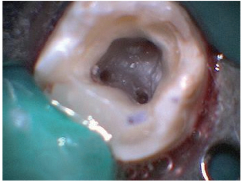
Figure 2 An intraoral photograph showing the wide buccolingual dimension of the mesial orifices.

Figure 3 (a) Transverse section cone-beam computed tomography (CBCT) scan image of tooth #47 clearly showing two separate mesial roots. Axial section of CBCT scan image (b) at the coronal third; (c) at the middle third; (d) at the apical third.
Reference
-
1. Ingle JI, Backland LK, Baumgarthner JC. Endodontics. In : Cleghorn BM, Goodacre CJ, Christie WH, editors. Morphology of teeth and their root canal system. 6th ed. Hamilton: BC Decker Inc.;2008. p. 151–220.2. Visser JB. Beitrag zur Kenntnis der menschlichen Zahnwurzelformen. Hilversum: Rotting;1948. p. 49–72.3. Ferraz JA, Pécora JD. Three-rooted mandibular molars in patients of Mongolian, Caucasian and Negro origin. Braz Dent J. 1993; 3:113–117.4. Slowey RR. Root canal anatomy. Road map to successful endodontics. Dent Clin North Am. 1979; 23:555–573.5. Sert S, Bayrl G. Taurodontism in six molars: a case report. J Endod. 2004; 30:601–602.
Article6. Fava LR, Weinfeld I, Fabri FP, Pais CR. Four second molars with single roots and single canals in the same patient. Int Endod J. 2000; 33:138–142.
Article7. Bond JL, Hartwell GR, Donnelly JC, Portell FR. Clinical management of middle mesial root canals in mandibular molars. J Endod. 1988; 14:312–314.
Article8. Kannan SK, Suganya , Santharam H. Supernumerary roots. Indian J Dent Res. 2002; 13:116–119.9. Peiris R, Pitakotuwage N, Takahashi M, Ohzeki S, Nakayama M, Sakurai S, Igarashi Y, Matsuno M, Sasaki K, Satake T, Kanazawa E. Mandibular permanent second molar with four roots and root canals: a case report. Odontology. 2009; 97:51–53.
Article10. Ravanshad S, Nabavizade MR. Endodontic treatment of a mandibular second molar with two mesial roots: report of a case. Iran Endod J. 2008; 3:137–140.11. Ahmed HMA, Luddin N. Accessory mesial roots and root canals in mandibular molar teeth: case reports, SEM analysis and literature review. Endod Pract Today. 2012; 6:195–205.12. Neville BW, Damm DD, Allen CM, Bouquot JE. Oral & Maxillofacial Pathology. In : Neville BW, Damm DD, Allen CM, Bouquot JE, editors. Abnormalities of teeth. 2nd ed. philadelphia: Saunders;2002. p. 88.13. Baert AL. Encyclopedia of diagnostic imaging. Volume 1. Seoul: Springer;2008. p. 427.14. Alt KW, Rösing FW, Teschler-Nicola M. Dental Anthropology. In : Türp JC, Alt KW, editors. Chapter 3.1. Anatomy and morphology of human teeth. Fundamentals, Limits and Prospects, Austria: Springer;1998. p. 71–94.15. Midtbø M, Halse A. Root length, crown height, and root morphology in Turner syndrome. Acta Odontol Scand. 1994; 52:303–314.
Article16. de Souza-Freitas JA, Lopes ES, Casati-Alvares L. Anatomic variations of lower first permanent molar roots in two ethnic groups. Oral Surg Oral Med Oral Pathol. 1971; 31:274–278.
Article17. Onda S, Minemura R, Masaki T, Funatsu S. Shape and number of the roots of the permanent molar teeth. Bull Tokyo Dent Coll. 1989; 30:221–231.18. Calberson FL, De Moor RJ, Deroose CA. The radix entomolaris and paramolaris: clinical approach in endodontics. J Endod. 2007; 33:58–63.
Article19. Jerome CE, Hanlon RJ Jr. Dental anatomical anomalies in Asians and Pacific Islanders. J Calif Dent Assoc. 2007; 35:631–636.20. Huang RY, Lin CD, Lee MS, Yeh CL, Shen EC, Chiang CY, Chiu HC, Fu E. Mandibular disto-lingual root: a consideration in periodontal therapy. J Periodontol. 2007; 78:1485–1490.
Article
- Full Text Links
-
- Actions
-
Cited
- CITED
-
- Close
- Share
-
- Similar articles
-
- Assessment of Root and Root Canal Morphology of Human Primary Molars using CBCT
- Observation of mandibular second molar roots and root canal morphology using dental cone-beam computed tomography
- A Study of Root Canals Morphology in Primary Molars using Computerized Tomography
- The characteristics of Korean multi-rooted teeth root trunk extracted by periodontal disease
- Evaluation of mesial root canal configuration of mandibular first molars using micro-computed tomography
