Prevalence and features of distolingual roots in mandibular molars analyzed by cone-beam computed tomography
- Affiliations
-
- 1Department of Conservative Dentistry, Wonkang University Daejeon Dental Hospital, Daejeon, Korea. profee@naver.com
- KMID: 2132934
- DOI: http://doi.org/10.5624/isd.2015.45.4.221
Abstract
- PURPOSE
This study evaluated the prevalence of distolingual roots in mandibular molars among Koreans, the root canal system associated with distolingual roots, and the concurrent appearance of a distolingual root in the mandibular first molar and a C-shaped canal in the mandibular second molar.
MATERIALS AND METHODS
Cone-beam computed tomographic images of 264 patients were screened and examined. Axial sections of 1056 mandibular molars were evaluated to determine the number of roots. The interorifice distances from the distolingual canal to the distobuccal canal were also estimated. Using an image analysis program, the root canal curvature was calculated. Pearson's chi-square test, the paired t-test, one-way analysis of variance, and post-hoc analysis were performed.
RESULTS
Distolingual roots were observed in 26.1% of the subjects. In cases where a distolingual root was observed in the mandibular molar, a significant difference was observed in the root canal curvature between the buccolingual and mesiodistal orientations. The maximum root canal curvature was most commonly observed in the mesiodistal orientation in the coronal portion, but in the apical portion, maximum root canal curvature was most often observed in the buccolingual orientation.
CONCLUSION
The canal curvature of distolingual roots was found to be very complex, with a different direction in each portion. No correlation was found between the presence of a distolingual root in the mandibular first molar and the presence of a C-shaped canal in the mandibular second molar.
Figure
-
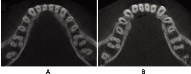
Fig. 1 Mandibular molars with a distolingual root visible in the axial section. A. Bilateral occurrence, B. Unilateral occurrence.
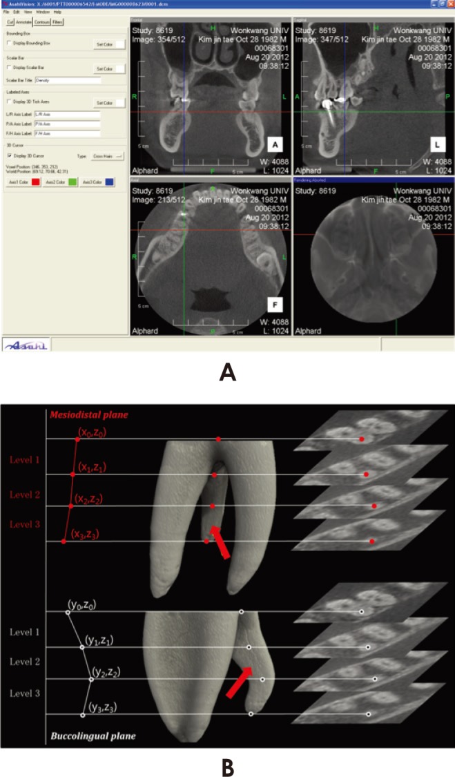
Fig. 2 A. Cone-beam computed tomographic images are displayed in three planes. The software automatically displays the locations of the designated points using three-dimensional coordinates (x, y, and z). B. The schema for the current study design. Red arrows indicate the distolingual roots of the mandibular molars. The centers of the root canals are plotted manually, and individual points are connected to create curves.
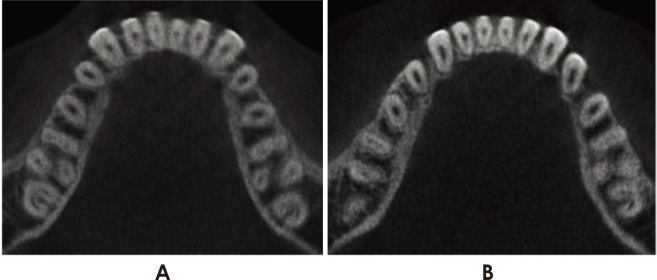
Fig. 3 Mandibular molars with the concurrent presence of a distolingual root and a C-shaped canal. A. Bilateral occurrence, B. Unilateral occurrence.
Cited by 1 articles
-
CBCT study of mandibular first molars with a distolingual root in Koreans
Hee-Ho Kim, Hyoung-Hoon Jo, Jeong-Bum Min, Ho-Keel Hwang
Restor Dent Endod. 2018;43(3):. doi: 10.5395/rde.2018.43.e33.
Reference
-
1. Yu DC, Schilder H. Cleaning and shaping the apical third of a root canal system. Gen Dent. 2001; 49:266–270. PMID: 12004725.2. de Pablo OV, Estevez R, Péix Sánchez M, Heilborn C, Cohenca N. Root anatomy and canal configuration of the permanent mandibular first molar: a systematic review. J Endod. 2010; 36:1919–1931. PMID: 21092807.
Article3. Carlsen O, Alexandersen V. Radix entomolaris: identification and morphology. Scand J Dent Res. 1990; 98:363–373. PMID: 2293344.
Article4. De Moor RJ, Deroose CA, Calberson FL. The radix entomolaris in mandibular first molars: an endodontic challenge. Int Endod J. 2004; 37:789–799. PMID: 15479262.
Article5. Calberson FL, De Moor RJ, Deroose CA. The radix entomolaris and paramolaris: clinical approach in endodontics. J Endod. 2007; 33:58–63. PMID: 17185133.
Article6. Gu Y, Lu Q, Wang P, Ni L. Root canal morphology of permanent three-rooted mandibular first molars: part II - measurement of root canal curvatures. J Endod. 2010; 36:1341–1346. PMID: 20647093.7. Abella F, Patel S, Durán-Sindreu F, Mercadé M, Roig M. Mandibular first molars with disto-lingual roots: review and clinical management. Int Endod J. 2012; 45:963–978. PMID: 22681628.
Article8. Gu Y, Lu Q, Wang H, Ding Y, Wang P, Ni L. Root canal morphology of permanent three-rooted mandibular first molars - part I: pulp floor and root canal system. J Endod. 2010; 36:990–994. PMID: 20478452.9. Nagaveni NB, Umashankar KV. Radix entomolaris in permanent mandibular first molars. Gen Dent. 2009; 57:e25–e29. PMID: 21467000.10. Jerome CE, Hanlon RJ Jr. Dental anatomical anomalies in Asians and Pacific Islanders. J Calif Dent Assoc. 2007; 35:631–636. PMID: 17993215.11. Gu Y, Zhou P, Ding Y, Wang P, Ni L. Root canal morphology of permanent three rooted mandibular first molars: part III - an odontometric analysis. J Endod. 2011; 37:485–490. PMID: 21419295.12. Seo MS, Park DS. C-shaped root canals of mandibular second molars in a Korean population: clinical observation and in vitro analysis. Int Endod J. 2004; 37:139–144. PMID: 14871181.13. Song JS, Choi HJ, Jung IY, Jung HS, Kim SO. The prevalence and morphologic classification of distolingual roots in the mandibular molars in a Korean population. J Endod. 2010; 36:653–657. PMID: 20307739.
Article14. Park JB, Kim N, Park S, Kim Y, Ko Y. Evaluation of root anatomy of permanent mandibular premolars and molars in a Korean population with cone-beam computed tomography. Eur J Dent. 2013; 7:94–101. PMID: 23407684.15. Song JS, Kim SO, Choi BJ, Choi HJ, Son HK, Lee JH. Incidence and relationship of an additional root in the mandibular first permanent molar and primary molars. Oral Surg Oral Med Oral Pathol Oral Radiol Endod. 2009; 107:e56–e60. PMID: 19101484.
Article16. Kim SY, Kim BS, Woo J, Kim Y. Morphology of mandibular first molars analyzed by cone-beam computed tomography in a Korean population: variations in the number of roots and canals. J Endod. 2013; 39:1516–1521. PMID: 24238439.
Article17. Sperber GH, Moreau JL. Study of the number of roots and canals in Senegalese first permanent mandibular molars. Int Endod J. 1998; 31:117–122. PMID: 9868938.
Article18. Onda S, Minemura R, Masaki T, Funatsu S. Shape and number of the roots of the permanent molar teeth. Bull Tokyo Dent Coll. 1989; 30:221–231. PMID: 2640921.19. Tu MG, Tsai CC, Jou MJ, Chen WL, Chang YF, Chen SY, et al. Prevalence of three-rooted mandibular first molars among Taiwanese individuals. J Endod. 2007; 33:1163–1166. PMID: 17889682.
Article20. Loh HS. Incidence and features of three-rooted permanent mandibular molars. Aust Dent J. 1990; 35:434–437. PMID: 2073191.
Article21. Curzon ME. Three-rooted mandibular permanent molars in English Caucasians. J Dent Res. 1973; 52:181. PMID: 4509495.
Article
- Full Text Links
-
- Actions
-
Cited
- CITED
-
- Close
- Share
-
- Similar articles
-
- CBCT study of mandibular first molars with a distolingual root in Koreans
- Observation of mandibular second molar roots and root canal morphology using dental cone-beam computed tomography
- Positional Relationship of the Mandibular Canal and Impacted Third Molars by Using Dental Cone Beam Computed Tomography
- Analysis and evaluation of relative positions of mandibular third molar and mandibular canal impacts
- Comparison of cone beam computed tomography and conventional panoramic radiography in assessing the topographic relationship between the mandibular canal and impacted third molars
