J Korean Assoc Oral Maxillofac Surg.
2014 Dec;40(6):278-284. 10.5125/jkaoms.2014.40.6.278.
Analysis and evaluation of relative positions of mandibular third molar and mandibular canal impacts
- Affiliations
-
- 1Department of Oral and Maxillofacial Surgery, College of Dentistry, Dankook University, Cheonan, Korea. Lee201@dku.edu
- KMID: 1799561
- DOI: http://doi.org/10.5125/jkaoms.2014.40.6.278
Abstract
OBJECTIVES
This study used cone-beam computed tomography (CBCT) images to categorize the relationships between the mandibular canal and the roots and investigated the prevalence of nerve damage.
MATERIALS AND METHODS
Through CBCT images, contact and three-dimensional positional relationships between the roots of the mandibular third molar and the mandibular canal were investigated. With this data, prevalence of nerve damage according to the presence of contact and three-dimensional positional relationships was studied. Other factors that affected the prevalence of nerve damage were also investigated.
RESULTS
When the mandibular third molar and the mandibular canal were shown to have direct contact in CBCT images, the prevalence of nerve damage was higher than in other cases. Also, in cases where the mandibular canal was horizontally lingual to the mandibular third molar and the mandibular canal was vertically at the cervical level of the mandibular third molar, the prevalence of nerve damage was higher than in opposite cases. The percentage of mandibular canal contact with the roots of the mandibular third molar was higher when the mandibular canal was horizontally lingual to the mandibular third molar. Finally, the prevalence of nerve damage was higher when the diameter of the mandibular canal lumen suddenly decreased at the contact area between the mandibular canal and the roots, as shown in CBCT images.
CONCLUSION
The three-dimensional relationship of the mandibular third molar and the mandibular canal can help predict nerve damage and can guide patient expectations of the possibility and extent of nerve damage.
Keyword
Figure
-
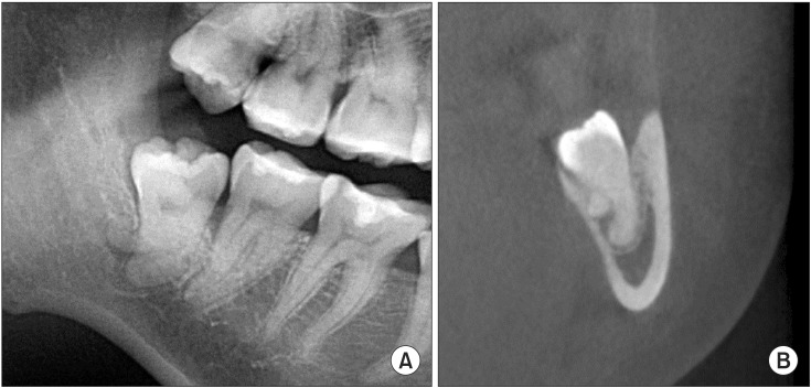
Fig. 1 Radiographic images of a patient with paresthesia. A. A panoramic radiographic image shows the roots of the mandibular third molar overlapping the mandibular canal. B. The roots show lingual bending, and the continuity of the canal of the inferior alveolar nerve is lost.
Reference
-
1. Robinson PP, Loescher AR, Yates JM, Smith KG. Current management of damage to the inferior alveolar and lingual nerves as a result of removal of third molars. Br J Oral Maxillofac Surg. 2004; 42:285–292. PMID: 15225944.2. Kipp DP, Goldstein BH, Weiss WW Jr. Dysesthesia after mandibular third molar surgery: a retrospective study and analysis of 1,377 surgical procedures. J Am Dent Assoc. 1980; 100:185–192. PMID: 6928147.
Article3. Cheung LK, Leung YY, Chow LK, Wong MC, Chan EK, Fok YH. Incidence of neurosensory deficits and recovery after lower third molar surgery: a prospective clinical study of 4338 cases. Int J Oral Maxillofac Surg. 2010; 39:320–326. PMID: 20061121.
Article4. Lopes V, Mumenya R, Feinmann C, Harris M. Third molar surgery: an audit of the indications for surgery, post-operative complaints and patient satisfaction. Br J Oral Maxillofac Surg. 1995; 33:33–35. PMID: 7718526.
Article5. Monaco G, Montevecchi M, Bonetti GA, Gatto MR, Checchi L. Reliability of panoramic radiography in evaluating the topographic relationship between the mandibular canal and impacted third molars. J Am Dent Assoc. 2004; 135:312–318. PMID: 15058618.
Article6. Gülicher D, Gerlach KL. Sensory impairment of the lingual and inferior alveolar nerves following removal of impacted mandibular third molars. Int J Oral Maxillofac Surg. 2001; 30:306–312. PMID: 11518353.
Article7. Jerjes W, Upile T, Shah P, Nhembe F, Gudka D, Kafas P, et al. Risk factors associated with injury to the inferior alveolar and lingual nerves following third molar surgery-revisited. Oral Surg Oral Med Oral Pathol Oral Radiol Endod. 2010; 109:335–345. PMID: 20097103.
Article8. Tantanapornkul W, Okouchi K, Fujiwara Y, Yamashiro M, Maruoka Y, Ohbayashi N, et al. A comparative study of cone-beam computed tomography and conventional panoramic radiography in assessing the topographic relationship between the mandibular canal and impacted third molars. Oral Surg Oral Med Oral Pathol Oral Radiol Endod. 2007; 103:253–259. PMID: 17234544.
Article9. Suomalainen A, Ventä I, Mattila M, Turtola L, Vehmas T, Peltola JS. Reliability of CBCT and other radiographic methods in preoperative evaluation of lower third molars. Oral Surg Oral Med Oral Pathol Oral Radiol Endod. 2010; 109:276–284. PMID: 20123411.
Article10. Rood JP, Shehab BA. The radiological prediction of inferior alveolar nerve injury during third molar surgery. Br J Oral Maxillofac Surg. 1990; 28:20–25. PMID: 2322523.
Article11. Flygare L, Ohman A. Preoperative imaging procedures for lower wisdom teeth removal. Clin Oral Investig. 2008; 12:291–302.
Article12. Susarla SM, Sidhu HK, Avery LL, Dodson TB. Does computed tomographic assessment of inferior alveolar canal cortical integrity predict nerve exposure during third molar surgery? J Oral Maxillofac Surg. 2010; 68:1296–1303. PMID: 20356665.
Article13. Susarla SM, Dodson TB. Preoperative computed tomography imaging in the management of impacted mandibular third molars. J Oral Maxillofac Surg. 2007; 65:83–88. PMID: 17174769.
Article14. Ohman A, Kivijärvi K, Blombäck U, Flygare L. Pre-operative radiographic evaluation of lower third molars with computed tomography. Dentomaxillofac Radiol. 2006; 35:30–35. PMID: 16421261.15. Mahasantipiya PM, Savage NW, Monsour PA, Wilson RJ. Narrowing of the inferior dental canal in relation to the lower third molars. Dentomaxillofac Radiol. 2005; 34:154–163. PMID: 15897286.
Article16. Kaeppler G. Conventional cross-sectional tomographic evaluation of mandibular third molars. Quintessence Int. 2000; 31:49–56. PMID: 11203906.17. Maegawa H, Sano K, Kitagawa Y, Ogasawara T, Miyauchi K, Sekine J, et al. Preoperative assessment of the relationship between the mandibular third molar and the mandibular canal by axial computed tomography with coronal and sagittal reconstruction. Oral Surg Oral Med Oral Pathol Oral Radiol Endod. 2003; 96:639–646. PMID: 14600702.
Article18. Nakamori K, Fujiwara K, Miyazaki A, Tomihara K, Tsuji M, Nakai M, et al. Clinical assessment of the relationship between the third molar and the inferior alveolar canal using panoramic images and computed tomography. J Oral Maxillofac Surg. 2008; 66:2308–2313. PMID: 18940497.
Article19. Eyrich G, Seifert B, Matthews F, Matthiessen U, Heusser CK, Kruse AL, et al. 3-Dimensional imaging for lower third molars: is there an implication for surgical removal? J Oral Maxillofac Surg. 2011; 69:1867–1872. PMID: 21419547.
Article20. Jhamb A, Dolas RS, Pandilwar PK, Mohanty S. Comparative efficacy of spiral computed tomography and orthopantomography in preoperative detection of relation of inferior alveolar neurovascular bundle to the impacted mandibular third molar. J Oral Maxillofac Surg. 2009; 67:58–66. PMID: 19070749.
Article
- Full Text Links
-
- Actions
-
Cited
- CITED
-
- Close
- Share
-
- Similar articles
-
- Radiographic evaluation of the course and visibility of the mandibular canal
- The clinical study of the mandibular canal location in mandibular molar areas using dentascan
- Assessment of the relationship between the mandibular third molar and the mandibular canal using panoramic radiograph and cone beam computed tomography
- Anatomical position of the mandibular canal in relation to the buccal cortical bone: relevance to sagittal split osteotomy
- Positional relationship between mandibular third molar and mandibular canal in cone beam computed tomographs
