J Korean Assoc Oral Maxillofac Surg.
2018 Aug;44(4):167-173. 10.5125/jkaoms.2018.44.4.167.
Anatomical position of the mandibular canal in relation to the buccal cortical bone: relevance to sagittal split osteotomy
- Affiliations
-
- 1Department of Oral and Maxillofacial Surgery, College of Dentistry, Dankook University, Cheonan, Korea. hanimplant@dankook.ac.kr
- KMID: 2419232
- DOI: http://doi.org/10.5125/jkaoms.2018.44.4.167
Abstract
OBJECTIVES
Classification of the degree of postoperative nerve damage according to contact with the mandibular canal and buccal cortical bone has been studied, but there is a lack of research on the difference in postoperative courses according to contact with buccal cortical bone. In this study, we divided patients into groups according to contact between the mandibular canal and the buccal cortical bone, and we compared the position of the mandibular canal in the second and first molar areas.
MATERIALS AND METHODS
Class III patients who visited the Dankook University Dental Hospital were included in this study. The following measurements were made at the second and first molar positions: (1) length between the outer margin of the mandibular canal and the buccal cortical margin (a); (2) mandibular thickness at the same level (b); (3) Buccolingual ratio=(a)/(b)×100; and (4) length between the inferior margin of the mandibular canal and the inferior cortical margin.
RESULTS
The distances from the canal to the buccal bone and from the canal to the inferior bone and mandibular thickness were significantly larger in Group II than in Group I. The buccolingual ratio of the canal was larger in Group II in the second molar region.
CONCLUSION
If mandibular canal is in contact with the buccal cortical bone, the canal will run closer to the buccal bone and the inferior border of the mandible in the second and first molar regions.
Figure
-
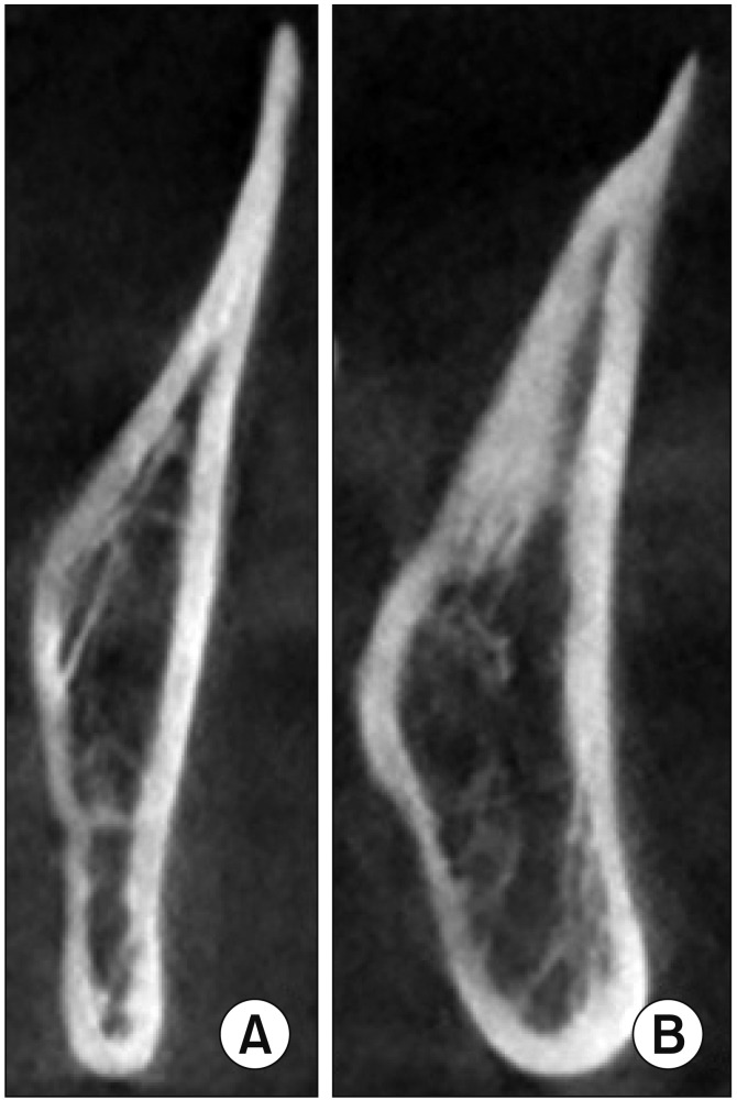
Fig. 1 A. Contact group. B. Seperate group.
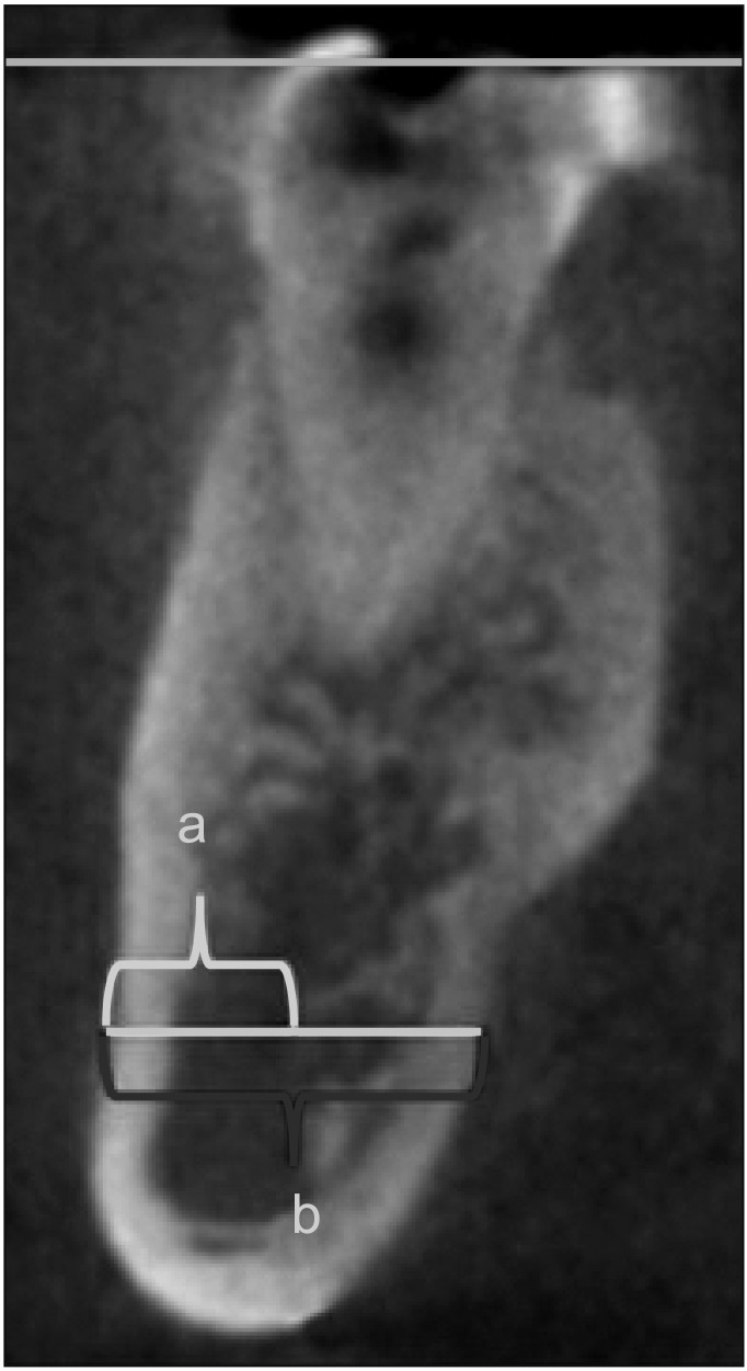
Fig. 2 Blue line (upper) shows occlusal plane. Yellow line is parallel to occlusal plane. ‘a’ is lengths between canal and buccal cortical bone. ‘b’ is mandible thickness at the same level.
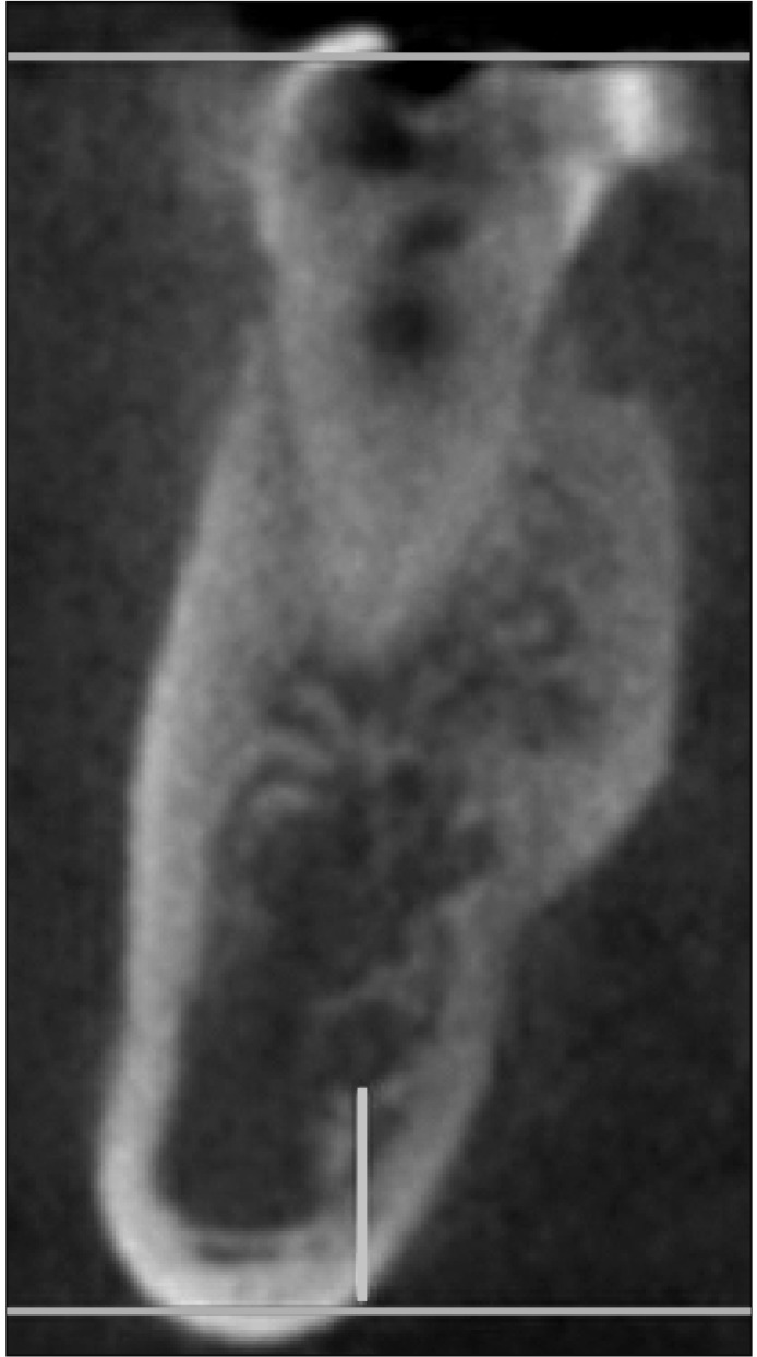
Fig. 3 Blue line (below) is parallel to occlusal plane. Yellow line is perpendicular to blue line. Lengths of yellow line is lengths between canal and inferior cortical bone.
Reference
-
1. Kawashima Y, Sakai O, Shosho D, Kaneda T, Gohel A. Proximity of the mandibular canal to teeth and cortical bone. J Endod. 2016; 42:221–224. PMID: 26725176.
Article2. Sato I, Ueno R, Kawai T, Yosue T. Rare courses of the mandibular canal in the molar regions of the human mandible: a cadaveric study. Okajimas Folia Anat Jpn. 2005; 82:95–101. PMID: 16350422.
Article3. Yamada T, Ishihama K, Yasuda K, Hasumi-Nakayama Y, Ito K, Yamaoka M, et al. Inferior alveolar nerve canal and branches detected with dental cone beam computed tomography in lower third molar region. J Oral Maxillofac Surg. 2011; 69:1278–1282. PMID: 21256640.
Article4. Xu GZ, Yang C, Fan XD, Yu CQ, Cai XY, Wang Y, et al. Anatomic relationship between impacted third mandibular molar and the mandibular canal as the risk factor of inferior alveolar nerve injury. Br J Oral Maxillofac Surg. 2013; 51:e215–e219. PMID: 23411471.
Article5. Yu IH, Wong YK. Evaluation of mandibular anatomy related to sagittal split ramus osteotomy using 3-dimensional computed tomography scan images. Int J Oral Maxillofac Surg. 2008; 37:521–528. PMID: 18450425.
Article6. Westermark A, Bystedt H, von Konow L. Inferior alveolar nerve function after mandibular osteotomies. Br J Oral Maxillofac Surg. 1998; 36:425–428. PMID: 9881783.
Article7. Westermark A, Bystedt H, von Konow L. Inferior alveolar nerve function after sagittal split osteotomy of the mandible: correlation with degree of intraoperative nerve encounter and other variables in 496 operations. Br J Oral Maxillofac Surg. 1998; 36:429–433. PMID: 9881784.
Article8. Yoshida T, Nagamine T, Kobayashi T, Michimi N, Nakajima T, Sasakura H, et al. Impairment of the inferior alveolar nerve after sagittal split osteotomy. J Craniomaxillofac Surg. 1989; 17:271–277. PMID: 2671043.
Article9. Lee CH, Lee BS, Choi BJ, Lee JW, Ohe JY, Yoo HY, et al. Recovery of inferior alveolar nerve injury after bilateral sagittal split ramus osteotomy (BSSRO): a retrospective study. Maxillofac Plast Reconstr Surg. 2016; 38:25. PMID: 27441184.
Article10. Wittwer G, Adeyemo WL, Beinemann J, Juergens P. Evaluation of risk of injury to the inferior alveolar nerve with classical sagittal split osteotomy technique and proposed alternative surgical techniques using computer-assisted surgery. Int J Oral Maxillofac Surg. 2012; 41:79–86. PMID: 21925838.
Article11. Panula K, Finne K, Oikarinen K. Neurosensory deficits after bilateral sagittal split ramus osteotomy of the mandible--influence of soft tissue handling medial to the ascending ramus. Int J Oral Maxillofac Surg. 2004; 33:543–548. PMID: 15308252.
Article12. Plooij JM, Naphausen MT, Maal TJ, Xi T, Rangel FA, Swennnen G, et al. 3D evaluation of the lingual fracture line after a bilateral sagittal split osteotomy of the mandible. Int J Oral Maxillofac Surg. 2009; 38:1244–1249. PMID: 19713076.
Article13. Muto T, Takahashi M, Akizuki K. Evaluation of the mandibular ramus fracture line after sagittal split ramus osteotomy using 3-dimensional computed tomography. J Oral Maxillofac Surg. 2012; 70:e648–e652. PMID: 23078827.
Article14. Rich J, Golden BA, Phillips C. Systematic review of preoperative mandibular canal position as it relates to postoperative neurosensory disturbance following the sagittal split ramus osteotomy. Int J Oral Maxillofac Surg. 2014; 43:1076–1081. PMID: 24837554.
Article15. Tsuji Y, Muto T, Kawakami J, Takeda S. Computed tomographic analysis of the position and course of the mandibular canal: relevance to the sagittal split ramus osteotomy. Int J Oral Maxillofac Surg. 2005; 34:243–246. PMID: 15741030.
Article16. Yamamoto R, Nakamura A, Ohno K, Michi KI. Relationship of the mandibular canal to the lateral cortex of the mandibular ramus as a factor in the development of neurosensory disturbance after bilateral sagittal split osteotomy. J Oral Maxillofac Surg. 2002; 60:490–495. PMID: 11988921.
Article17. Fridrich KL, Holton TJ, Pansegrau KJ, Buckley MJ. Neurosensory recovery following the mandibular bilateral sagittal split osteotomy. J Oral Maxillofac Surg. 1995; 53:1300–1306. discussion 1306-7. PMID: 7562195.
Article18. Kim HJ, Lee HY, Chung IH, Cha IH, Yi CK. Mandibular anatomy related to sagittal split ramus osteotomy in Koreans. Yonsei Med J. 1997; 38:19–25. PMID: 9100479.
Article19. Martone CH, Ben-Josef AM, Wolf SM, Mintz SM. Dimorphic study of surgical anatomic landmarks of the lateral ramus of the mandible. Oral Surg Oral Med Oral Pathol. 1993; 75:436–438. PMID: 8464606.
Article20. Pogrel MA, Schmidt BL, Ammar A. The presence of the antilingula and its relationship to the true lingula. Br J Oral Maxillofac Surg. 1995; 33:235–238. PMID: 8736750.
Article21. Park HS, Lee JH. A comparative study on the location of the mandibular foramen in CBCT of normal occlusion and skeletal class II and III malocclusion. Maxillofac Plast Reconstr Surg. 2015; 37:25. PMID: 26301208.
Article22. Zhou C, Jeon TH, Jun SH, Kwon JJ. Evaluation of mandibular lingula and foramen location using 3-dimensional mandible models reconstructed by cone-beam computed tomography. Maxillofac Plast Reconstr Surg. 2017; 39:30. PMID: 29109943.
Article23. Nasel CJ, Pretterklieber M, Gahleitner A, Czerny C, Breitenseher M, Imhof H. Osteometry of the mandible performed using dental MR imaging. AJNR Am J Neuroradiol. 1999; 20:1221–1227. PMID: 10472975.24. Levine MH, Goddard AL, Dodson TB. Inferior alveolar nerve canal position: a clinical and radiographic study. J Oral Maxillofac Surg. 2007; 65:470–474. PMID: 17307595.
Article25. Promma L, Sakulsak N, Putiwat P, Amarttayakong P, Iamsaard S, Trakulsuk H, et al. Cortical bone thickness of the mandibular canal and implications for bilateral sagittal split osteotomy: a cadaveric study. Int J Oral Maxillofac Surg. 2017; 46:572–577. PMID: 28089388.
Article26. Nagadia R, Tay AB, Chan LL, Chan ES. The spatial location of the mandibular canal in Chinese: a CT study. Int J Oral Maxillofac Surg. 2011; 40:1401–1405. PMID: 21862289.
Article27. Yoshioka I, Tanaka T, Khanal A, Habu M, Kito S, Kodama M, et al. Relationship between inferior alveolar nerve canal position at mandibular second molar in patients with prognathism and possible occurrence of neurosensory disturbance after sagittal split ramus osteotomy. J Oral Maxillofac Surg. 2010; 68:3022–3027. PMID: 20739116.
Article
- Full Text Links
-
- Actions
-
Cited
- CITED
-
- Close
- Share
-
- Similar articles
-
- Study On The Relationship Of The Inferior Alveolar Nerve Position Between Buccal And Lingual Side Using Ct And Orthpantomogram
- Mandibular anatomy related to sagittal split ramus osteotomy in Koreans
- Bifid Mandibular Canal: Radiographic Observation and Clinical Relevance: A Case Report
- Skeletal relapse pattern after sagittal split ramus osteotomy of mandibular prognathic patient
- Bone changes after bilateral sagittal split osteotomy for mandibular prognathism
