Primary Epidural Peripheral Primitive Neuroectodermal Tumor of the Lumbar Spine: A Case Report
- Affiliations
-
- 1Department of Radiology, Hanyang University Hospital, Seoul, Korea. radsh@medimail.co.kr
- 2Department of Pathology, Hanyang University Hospital, Seoul, Korea.
- 3Department of Pediatrics, Hanyang University Hospital, Seoul, Korea.
- KMID: 2040810
- DOI: http://doi.org/10.3348/jksr.2011.64.5.435
Abstract
- A primitive neuroectodermal tumor (PNET) is a highly malignant tumor in children and young adults, and extremely rare in the spine. We report a case of a primary epidural peripheral PNET of the lumbar spine. The present extremely rare case of primary epidural peripheral PNET of the lumbar spine illustrates the unexpected occurrence and should be included in differential diagnoses for patients with spinal tumors.
MeSH Terms
Figure
-

Fig. 1 Primary epidural peripheral primitive neuroectodermal tumor in a 12-year-old boy with low back pain. Axial MR images demonstrate an extradural mass with multi-lobulating contour, extending from the L1 to L2 level with spinal cord compression and extending to the right neural foraminal zone and paraspinal muscle. A. Axial T2WI (TR/TE: 4000/123) demonstrates an isointensity, with multifocal hyperintensity which appear as a cyst-like lesion. B. T1WI (TR/TE: 600/7) shows isointensity of the epidural tumor. C. Gadolinium-enhanced fat suppressed T1WI shows mild enhancement.
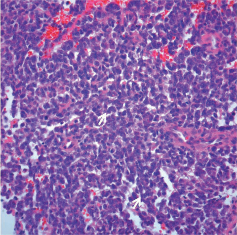
Fig. 2 Photomicrographs of the primary epidural pPNET. The tumor is composed of small round cells with scanty cytoplasm and a vague rossette-like arrangement (arrows) (hematoxylin & eosin staining, ×400).
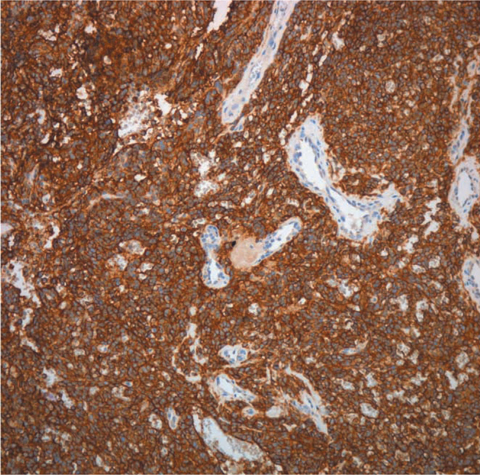
Fig. 3 On immunohistochemical staining, the tumor cells showed diffuse, strong membranous positivity for CD99 (×100).
Reference
-
1. Deme S, Ang LC, Skaf G, Rowed DW. Primary intramedullary primitive neuroectodermal tumor of the spinal cord: case report and review of literature. Neurosurgery. 1997; 41:1417–1420.2. Chang SI, Tsai MC, Tsai MD. An unusual primitive neuroectodermal tumor in the thoracic epidural space. J Clin Neurosci. 2010; 17:261–263.3. Kumar R, Reddy SJ, Wani AA, Pal L. Primary spinal primitive neuroectodermal tumor: case series and reviews of the literature. Pediatr Neurosurg. 2007; 43:1–6.4. Kiatsoontorn K, Takami T, Ichinose T, Chokyu I, Tsuyuguchi N, Ohsawa M, et al. Primary epidural peripheral primitive neuroectodermal tumor of the thoracic spine. Neurol Med Chir (Tokyo). 2009; 49:542–545.5. Kim YW, Jin BH, Kim TS, Cho YE. Primary intraspinal primitive neuroectodermal tumor at conus medullaris. Yonsei Med J. 2004; 45:533–538.6. Hart MN, Earle KM. Primitive neuroectodermal tumors of the brain in children. Cancer. 1973; 32:890–897.7. Dorfmüller G, Würtz FG, Umschaden HW, Kleinert R, Ambros PF. Intraspinal primitive neuroectodermal tumour: report of two cases and review of the literature. Acta Neurochir. 1999; 141:1169–1175.8. Ambros IM, Ambros PF, Strehl S, Kovar H, Gadner H, Salzer-Kuntschik M. MIC2 is a specific marker for Ewing's sarcoma and peripheral primitive neuroectodermal tumors: evidence for a common histogenesis of Ewing's sarcoma and peripheral primitive neuroectodermal tumors from MIC2 expression and specific chromosome aberration. Cancer. 1991; 67:1886–1893.9. Li MJ, Pan ST, Cheng C. MRI of primary spinal epidural primitive neuroectodermal tumor: a case report. Chin J Radiol. 2008; 33:91–95.10. Shin JH, Lee HK, Rhim SC, Cho KJ, Choi CG, Suh DC. Spinal epidural extraskeletal Ewing sarcoma: MR findings in two cases. AJNR Am J Neuroradiol. 2001; 22:795–798.
- Full Text Links
-
- Actions
-
Cited
- CITED
-
- Close
- Share
-
- Similar articles
-
- Primitive Neuroectodermal Tumor of Mediastinum in an Adult: A Case Report
- Peripheral Primitive Neuroectodermal Tumor of the Stomach: A Case Report
- Ewing Sarcoma/Peripheral Primitive Neuroectodermal Tumor in an Adolscence, Manifested as Isolated Cervical Mass
- A Case of Primitive Neuroectodermal Tumor in the Maxillary Sinus
- Peripheral Neuroblastoma: A Report of One Case
