J Korean Soc Radiol.
2010 Sep;63(3):235-238. 10.3348/jksr.2010.63.3.235.
Peripheral Primitive Neuroectodermal Tumor of the Stomach: A Case Report
- Affiliations
-
- 1Department of Radiology, College of Medicine, Chungnam National University, Korea. jscho@cnu.ac.kr
- 2Department of Internal Medicine, College of Medicine, Chungnam National University, Korea.
- 3Department of Surgery, College of Medicine, Chungnam National University, Korea.
- 4Department of Pathology, College of Medicine, Chungnam National University, Korea.
- KMID: 2097898
- DOI: http://doi.org/10.3348/jksr.2010.63.3.235
Abstract
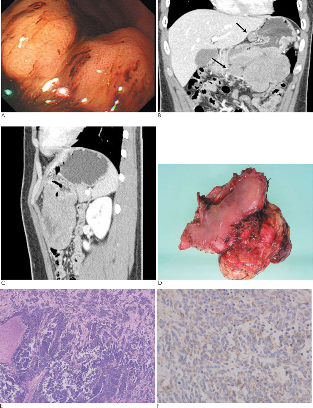
- Peripheral primitive neuroectodermal tumors (peripheral PNETs) are very rare and highly aggressive soft-tissue malignancies originating from the neural crest. To the best of our knowledge, only a few cases of peripheral PNETs of the stomach have been reported in the literature. We report a case of large peripheral primitive neuroectodermal tumor of the stomach with MDCT findings in a 22-year-old man presenting epigastric pain and vomiting.
MeSH Terms
Figure
Reference
-
1. Kimber C, Michalski A, Spitz L, Pierro A. Primitive neuroectodermal tumours: anatomic location, extent of surgery, and outcome. J Pediatr Surg. 1998; 33:39–41.2. Soulard R, Claude V, Camparo P, Dufau JP, Saint-Blancard P, Gros P. Primitive neuroectodermal tumor of the stomach. Arch Pathol Lab Med. 2005; 129:107–110.3. Katz RL, Quezado M, Senderowicz AM, Villalba L, Laskin WB, Tsokos M. An intra-abdominal small round cell neoplasm with features of primitive neuroectodermal and desmoplastic round cell tumor and a EWS/FLI-1 fusion transcript. Hum Pathol. 1997; 28:502–509.4. Inaba H, Ohta S, Nishimura T, Takamochi K, Ishida I, Etoh T, et al. An operative case of primitive neuroectodermal tumor in the posterior mediastinum. Kyobu Geka. 1998; 51:250–253.5. Amin HM, Candel AG, Husain AN. Pathologic quiz case. A 22-year-old man with an abdominal mass. Arch Pathol Lab Med. 1999; 123:541–543.6. Gasecki D, Izycka-Swieszewska E, Szymkiewicz-Rogowska A, Kopczynski S, Mechlinska-Baczkowska J. Primitive neuroectodermal tumor of rare localization in two members of one family. Neurol Neurochir Pol. 1999; 33:1415–1423.7. Ibarburen C, Haberman JJ, Zerhouni EA. Peripheral primitive neuroectodermal tumors. CT and MRI evaluation. Eur J Radiol. 1996; 21:225–232.8. Kim MS, Kim B, Park CS, Song SY, Lee EJ, Park NH, et al. Radiologic findings of peripheral primitive neuroectodermal tumor arising in the retroperitoneum. AJR Am J Roentgenol. 2006; 186:1125–1132.9. Peter M, Gilbert E, Delattre O. A multiplex real-time PCR assay for the detection of gene fusions observed in solid tumors. Lab Invest. 2001; 81:905–912.10. Ushigome S, Machinami R, Sorensen PH. In Ewing sarcoma/primitive neuroectodermal tumour (PNET). In : Fletcher CDM, Unnik K, Mertens F, editors. World Health Organization Classification of Tumours Pathology and Genetics of Tumours of Soft Tissue and Bone. Lyon, France: IARC Press;2002. p. 297–300.


