Korean Circ J.
2007 Aug;37(8):365-372. 10.4070/kcj.2007.37.8.365.
Prognostic Significance of Descending Thoracic Aorta Intima-Media Thickness in Patients with Coronary Atherosclerosis
- Affiliations
-
- 1Division of Cardiology, Heart Center, College of Medicine, Konyang University, Daejeon, Korea. janghobae@yahoo.co.kr
- KMID: 2029064
- DOI: http://doi.org/10.4070/kcj.2007.37.8.365
Abstract
-
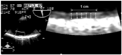
BACKGROUND AND OBJECTIVES: While the clinical significance of descending thoracic aorta intima-media thickness (DTA-IMT) remains unclear, common carotid IMT (CIMT) is known to be associated with major adverse cardiovascular events (MACE) in patients with atherosclerotic disease.
SUBJECTS AND METHODS
A total of 104 patients (mean age, 59 yrs; 69 male) with angiographically proven coronary atherosclerosis underwent transesophageal echocardiography (TEE) for DTA-IMT measurement and carotid scanning for CIMT measurement. The patients were divided into two groups based on the median IMT value, and they were followed up for cardiovascular events and all-cause mortality for a period of 50+/-21 months.
RESULTS
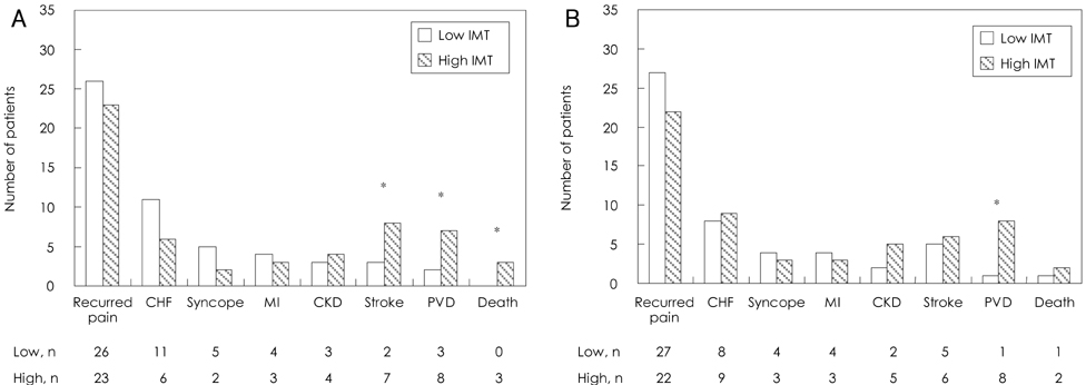
Patients having a higher DTA-IMT value (n=44, >2.1 mm) had a higher chance of stroke (6.7% vs. 2.8%, p=0.04), peripheral vascular disease (6.7% vs. 1.9%, p=0.02), and death (2.9% vs. 0%, p=0.04) than those who had lower DTA-IMT values (n=60, < or =2.1 mm). The patients who had higher CIMT values (n=49, >0.089 mm) had a higher chance of peripheral vascular disease (16% vs 2%, p=0.009) than those having lower IMT values (n=55, < or =0.089 mm). However, there was no significant difference between the groups in terms of recurrent chest pain, heart failure, syncope, myocardial infarction or chronic kidney disease during the follow-up period. Multivariate Cox regression analysis revealed that increased DTA-IMT was associated with stroke (OR, 4.29; 95% CI, 1.076-17.181; p=0.039) and peripheral vascular disease (OR, 9.37; 95% CI, 1.571-55.499; p=0.014), whereas increased CIMT was associated with peripheral vascular disease (OR, 14.365; 95% CI, 1.050-196.540; p=0.046).
CONCLUSION
This study suggests that descending thoracic aorta IMT is more closely associated with prognosis in patients with coronary atherosclerosis than CIMT.
Keyword
MeSH Terms
Figure
Cited by 1 articles
-
Differences of Aortic Stiffness and Aortic Intima-Media Thickness According to the Type of Initial Presentation in Patients with Ischemic Stroke
Hyun Ju Yoon, Kye Hun Kim, Sang Hyun Lee, Yi Rang Yim, Kyung Jin Lee, Keun Ho Park, Doo Sun Sim, Nam Sik Yoon, Young Joon Hong, Hyung Wook Park, Ju Han Kim, Youngkeun Ahn, Myung Ho Jeong, Jeong Gwan Cho, Jong Chun Park
J Cardiovasc Ultrasound. 2013;21(1):12-17. doi: 10.4250/jcu.2013.21.1.12.
Reference
-
1. Kuller L, Schemanski L, Psaty B, et al. Subclinical disease as an independent risk factor for cardiovascular disease. Circulation. 1995. 92:720–726.2. O'Leary DH, Polak JF, Wolfson SK Jr, et al. Use of sonography to evaluate carotid atherosclerosis in the elderly. Stroke. 1991. 22:1155–1163.3. Salonen R, Salonen JT. Determinants of carotid intima-media thickness: a population-based ultrasonography study in eastern Finnish men. J Intern Med. 1991. 229:225–231.4. Bots ML, Witterman JC, Grobbee DE. Carotid intima-media wall thickness in elderly women with and without atherosclerosis of the abdominal aorta. Atherosclerosis. 1993. 102:99–105.5. Bae JH, Kim KY. Impact of left ventricular ejection fraction on endothelial function and carotid Intima-media thickness in patients with coronary artery disease. Korean Circ J. 2005. 35:375–381.6. O'Leary DH, Polak JF, Kronmal RA, Manolio TA, Burke GL, Wolfson SK Jr. Carotid intima-media thickness as a risk factor for myocardial infarction and stroke in older adults. N Engl J Med. 1999. 340:14–22.7. O'Leary DH, Polak JF, Wolfson SK Jr, et al. Use of sonography to evaluate carotid atherosclerosis in the elderly. Stroke. 1991. 22:1155–1163.8. Grobbee D, Bots ML. Carotid artery intima-media thickness as a indicator of generalized atherosclerosis. J Intern Med. 1994. 236:567–573.9. Jeong IB, Bae JH, Kim KY, et al. The carotid intima-media thickness as a screening test for coronary artery disease. Korean Circ J. 2005. 35:460–466.10. Amarenco P, Cohen A, Tzourio C, et al. Atherosclerotic disease of the aortic arch and the risk of ischemic stroke. N Engl J Med. 1994. 331:1474–1479.11. Rohani M, Jogestrand T, Ekberg M, et al. Interrelation between the extent of atherosclerosis in the thoracic aorta, carotid intima-media thickness and the extent of coronary artery disease. Atherosclerosis. 2005. 179:311–316.12. Bae JH, Bassenge E, Park KR, Kim KY, Schwemmer M. Significance of the intima-media thickness of the thoracic aorta in patients with coronary atherosclerosis. Clin Cardiol. 2003. 26:574–578.13. Lehmann ED, Hopkins KD, Gosling RG. Atherosclerosis in the ascending aorta and risk of ischemic stroke. Lancet. 1995. 346:589–590.14. Matsumura Y, Takata J, Yabe T, Furuno T, Chikamori T, Doi YL. Atherosclerotic aortic plaque detected by transesophageal echocardiography: its significance and limitation as a marker for coronary artery disease in the elderly. Chest. 1997. 112:81–86.15. Agarwal R. Estimating GFR from serum creatinine concentration: pitfalls of GFR-estimating equations. Am J Kidney Dis. 2005. 45:610–613.16. Bostom AG, Kronrnberg F, Ritz E. Predictive performance of renal function equations for patients with chronic kidney disease and normal serum creatinine levels. J Am Soc Nephrol. 2002. 13:2140–2144.17. Pignoli P, Tremoli E, Poli A, Oreste P, Paoletti R. Intimal plus medial thickness of the arterial wall: a direct measurement with ultrasound imaging. Circulation. 1986. 74:1399–1406.18. Kallikazaros IE, Tsioufis CP, Stefanadis CI, Pitsavos CE, Toutouzas PK. Closed relation between carotid and ascending aortic atherosclerosis in cardiac patients. Circulation. 2000. 102:III263–III268.19. Rosvall M, Janzon L, Berglund G, Engstrom G, Hedblad B. Incidence of stroke is related to carotid IMT even in the absence of plaque. Atherosclerosis. 2005. 179:325–331.20. Fabris F, Zanocchhi M, Fonte G, et al. Carotid plaque, aging, and risk factors: a study of 457 subjects. Stroke. 1994. 25:1133–1140.21. Spence JD. Ultrsound measurement of carotid plaque as a surrogate outcome for coronary artery disease. Am J Cardiol. 2002. 89:10B–16B.22. Parfrey PS, Foley RN. The clinical epidemiology of cardiac disease in chronic renal failure. J Am Soc Nephrol. 1999. 10:1606–1615.23. Fried LF, Shlipak MG, Crump C, et al. Renal insufficiency as a predictor of cardiovascular outcomes and mortality in elderly individuals. J Am Coll Cardiol. 2003. 41:1364–1372.24. Culleton BF, Larson MG, Wilson PW, Evans JC, Parfrey PS, Levy D. Cardiovascular disease and mortality in a community-based cohort with mild renal insufficiency. Kidney Int. 1999. 56:2214–2219.
- Full Text Links
- Actions
-
Cited
- CITED
-
- Close
- Share
- Similar articles
-
- Transesophageal Echocardiographic Evaluation of Atherosclerosis
- Carotid ultrasound in patients with coronary artery disease
- Measurements of Carotid Intima, Media, and Intima-media Thickness and Their Clinical Importance
- Do we need individual measurement of carotid intima and media thickness?
- Penetrating Atherosclerotic Ulcer of the Descending Thoracic Aorta in a Patient with Heterozygote Familial Hypercholesterolemia


