J Korean Acad Prosthodont.
2009 Jan;47(1):12-20. 10.4047/jkap.2009.47.1.12.
Wettability of titanium implants depending upon surface properties
- Affiliations
-
- 1Advanced Prosthodontics, Graduate School of Clinical Dentistry, Korea University, Korea. swshin@korea.ac.kr
- KMID: 2000381
- DOI: http://doi.org/10.4047/jkap.2009.47.1.12
Abstract
- STATEMENT OF PROBLEM: When an implant is fixed, a fixture comes into contact with a tissue fluid. Adhesion of a tissue fluid to a surface of implant is various case by case.
PURPOSE: The ultimate goal of this work is to analyze a correlation between a surface roughness and wettability of implant specimens. A measurement for wettability is performed considering 4 types of specimen implant with surface treatments different from each other to investigate the change of wettability with the elapse of time.
MATERIAL AND METHODS: Firstly, 20 specimens of titanium were prepared. The specimen were made of a commercial Titanium Grade IV with the diameter of 10 mm and the thickness of 1 mm. According to the method of surface treatment, the specimens were classified into 4 groups of 5 specimens per group. Group A: Machined Surface Group B: Anodized surface Group C: RBM (HA blasting) surface Group D: CMP (calcium methaphosphate) coating surface. Surface roughness of specimen was measured using SV-3000S4 (Mituyoto, Japan). The measurement was based on the standard of JIS1994. Sessile drop method was used to measure the wettability, which measures contact angle between implant disc and saline with the time interval of 5, 10, and 15 seconds. SPSS 11.0 was used to analyze the collected data. In order to analyze the difference of wettability and surface roughness according to implant surface treatment method. The statistical significance was tested with the confidence level of 95 percent. Pearson's correlation coefficient was used to evaluate the correlation of surface roughness and wettability.
RESULTS
The difference of surface roughness was statistically significant in the order of Group C (1.69 +/- 0.26), Group D (1.58 +/- 0.16), Group B (0.78 +/- 0.14) Group A (0.18 +/- 0.05). The wettability has also a statistically significant difference, which was in the order of group B (17.70 +/- 2.66), Group C (27.86 +/- 4.52), Group D (66.28 +/- 3.70) Group A (70.52 +/- 8.00). There was no difference in wettability with the passage of time.
CONCLUSIONS
1. The surface roughness was high in the order of RBM, CMP, Anodized, Machined group (P < .05). 2. The wettability was high in the order of Anodized, RBM, CMP, Machined group (P < .05). 3. There was no statistical significance in the correlation of surface roughness and wettability.
Keyword
Figure
-

Fig. 1. Surface treated disc.
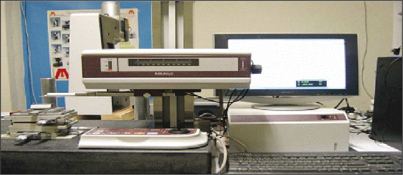
Fig. 2. Disc surface roughness measurement machine.
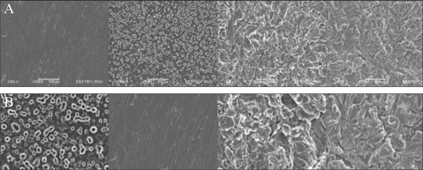
Fig. 3. SEM photographs of surface treated titanium. A (× 500), B (× 1,000)
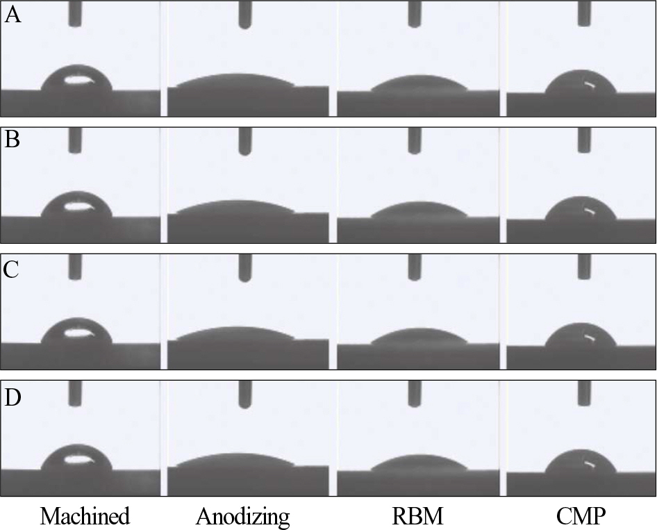
Fig. 4. Photographs of surface wettability. A, after 1 second B, after 2 second C, after 3 second D, after 4 second

Fig. 5. Change of surface wettability by the time processing. 1: Machined surface 2: Anodized surface 3: RBM surface 4: CMP surface.
Reference
-
1.Bra ° nemark PI., Adell R., Breine U., Hansson BO., Lindstrom J., Ohlsson A. Intra-osseous anchorage of dental prostheses. I. Experimental studies. Scand J Plast Reconstr Surg. 1969. 3:81–100.2.Adell R., Lekholm U., Rockler B., Bra ° nemark PI. A 15-year study of osseointegrated implants in the treatment of the edentulous jaw. Int J Oral Surg. 1981. 10:387–416.
Article3.Adell R., Eriksson B., Lekholm U., Bra ° nemark PI., Jemt T. Long-term follow-up study of osseointegrated implants in the treatment of totally edentulous jaws. Int J Oral Maxillofac Implants. 1990. 5:347–59.4.Albrektsson T., Dahl E., Enbom L., Engevall S., Engquist B., Eriksson AR., Feldmann G., Freiberg N., Glantz PO., Kjellman O, et al. Osseointegrated oral implants. A Swedish multicenter study of 8139 consecutively inserted Nobelpharma implants. J Periodontol. 1988. 59:287–96.5.Albrektsson T., Sennerby L. State of the art in oral implants. J Clin Periodontol. 1991. 18:474–81.
Article6.Eckert SE., Parein A., Myshin HL., Padilla JL. Validation of dental implant systems through a review of literature supplied by system manufacturers. J Prosthet Dent. 1997. 77:271–9.
Article7.Naert I., Quirynen M., van Steenberghe D., Darius P. A study of 589 consecutive implants supporting complete fixed prostheses. Part II: Prosthetic aspects. J Prosthet Dent. 1992. 68:949–56.
Article8.Cordioli G., Majzoub Z., Piattelli A., Scarano A. Removal torque and histomorphometric investigation of 4 different titanium surfaces: an experimental study in the rabbit tibia. Int J Oral Maxillofac Implants. 2000. 15:668–74.9.Gotfredsen K., Wennerberg A., Johansson C., Skovgaard LT., Hj фrting-Hansen E. Anchorage of TiO2-blasted, HA-coated, and machined implants: an experimental study with rabbits. J Biomed Mater Res. 1995. 29:1223–31.10.Gotfredsen K., Berglundh T., Lindhe J. Anchorage of titanium implants with different surface characteristics: an experimental study in rabbits. Clin Implant Dent Relat Res. 2000. 2:120–8.
Article11.Esposito M., Hirsch JM., Lekholm U., Thomsen P. Failure patterns of four osseointegrated oral implant systems. J Mater Sci Mater Med. 1997. 8:843–7.12.Sul YT., Johansson CB., Petronis S., Krozer A., Jeong Y., Wennerberg A., Albrektsson T. Characteristics of the surface oxides on turned and electrochemically oxidized pure titanium implants up to dielectric breakdown: the oxide thickness, micropore configurations, surface roughness, crystal structure and chemical composition. Biomaterials. 2002. 23:491–501.13.Buser D., Broggini N., Wieland M., Schenk RK., Denzer AJ., Cochran DL., Hoffmann B., Lussi A., Steinemann SG. Enhanced bone apposition to a chemically modified SLA titanium surface. J Dent Res. 2004. 83:529–33.
Article14.Park JY., Gemmell CH., Davies JE. Platelet interactions with titanium: modulation of platelet activity by surface topography. Biomaterials. 2001. 22:2671–82.
Article15.Kim JS., Shin SY., Ryu JJ. A study on the stability of 5 different surface treatment methods to dental implant using resonance frequency and histomorphometric analysis. J Korean Acad Prosthodont. 2005. 43:78–94.16.MacDonald DE., Deo N., Markovic B., Stranick M., Somasundaran P. Adsorption and dissolution behavior of human plasma fibronectin on thermally and chemically modified titanium dioxide particles. Biomaterials. 2002. 23:1269–79.
Article17.Rupp F., Scheideler L., Rehbein D., Axmann D., Geis-Gerstorfer J. Roughness induced dynamic changes of wettability of acid etched titanium implant modifications. Biomaterials. 2004. 25:1429–38.
Article18.Listgarten MA., Buser D., Steinemann SG., Donath K., Lang NP., Weber HP. Light and transmission electron microscopy of the intact interfaces between non-submerged titanium-coated epoxy resin implants and bone or gingiva. J Dent Res. 1992. 71:364–71.
Article19.Gotfredsen K., Nimb L., Hjorting-Hansen E., Jensen JS., Holmen A. Histomorphometric and removal torque analysis for TiO2-blasted titanium implants. An experimental study on dogs. Clin Oral Implants Res. 1992. 3:77–84.20.Buser D., Schenk RK., Steinemann S., Fiorellini JP., Fox CH., Stich H. Influence of surface characteristics on bone integration of titanium implants. A histomorphometric study in miniature pigs. J Biomed Mater Res. 1991. 25:889–902.
Article21.Wilke HJ., Claes L., Steinmann S. The influence of various titanium surfaces on the interface shear strength between implants and bone. Clinical implant Materials. Advances in Biomaterials. Elsevier Science Publishers B. V..22.Hanson S. On the role of surface roughness for load bearing bone implants M. SC. Thesis Centre for Biomechanics. Calmers University of Goteborg. Goteborg. 1991.
- Full Text Links
-
- Actions
-
Cited
- CITED
-
- Close
- Share
-
- Similar articles
-
- Effects of various surface treatments for titanium on surface micro roughness, static wettability, fibronectin adsorption
- The effects of saline soaking on the removal torque of titanium implants in rabbit tibia after 10 days
- A Biocompatibility Evaluation of Hydroxyapaite·Titania Surface for Dental Implant
- A comprehensive review of techniques for biofunctionalization of titanium
- Wettability and drug delivery of functionally graded nano-micro porous titanium surface
