Reestablishing the occlusal plane in full mouth rehabilitation patient, using Shilla system
- Affiliations
-
- 1Department of Prosthodontics, School of Dentistry, Chonnam National University, Gwangju, Korea. yhsdent@chonnam.ac.kr
- KMID: 2000184
- DOI: http://doi.org/10.4047/jkap.2013.51.1.33
Abstract
- Occlusal plane is a sagittal expression of dental arch form, and it composes the shape of occlusion, which is one of the most important elements of Maxillo-oral system. In this case, vertical, horizontal coordinates of bionic-median-sagittal plane was produced in articulator, and to achieve relation of left and right position of upper, lower teeth and deficits in alveola, Shilla system was used to reconstruct occlusal plane. In this case, a 41 year-old male patient visited for fracture of 10 unit metal-ceramic fixed partial denture of upper anterior teeth and for overall treatment. Clinical, radiographical, model examination was held, full mouth rehabilitation was achieved by placing dental implant. Maxillo-oral relation was recorded using Gothic arch Tracer complex and were mounted. And for the next step, we estimated original occlusal plane using Shilla system. After analysis we produced diagnosis wax pattern. On the basis of this, radiography stent was manufactured and dental implant was placed, and temporary prosthesis was made by using diagnosis wax pattern. Cross mounting and anterior guiding table were performed in order to reproduce temporary restoration morphology and bite pattern, followed by final restoration made of all ceramic crown with zirconia coping. As stated above, appropriately esthetic and functional results can be seen in using Shilla system in diagnosis and treatment procedure of full mouth rehabilitation patient.
MeSH Terms
Figure
-
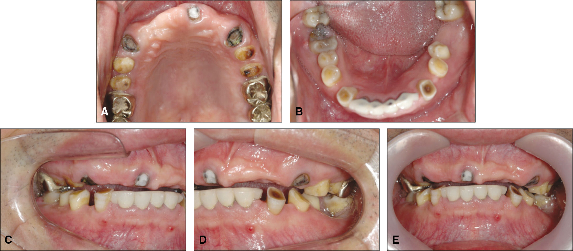
Fig. 1. Initial intraoral photograph. A: Maxillary occlusal view, B: Mandibular occlusal view, C: Right buccal view, D: Left buccal view, E: Frontal view.
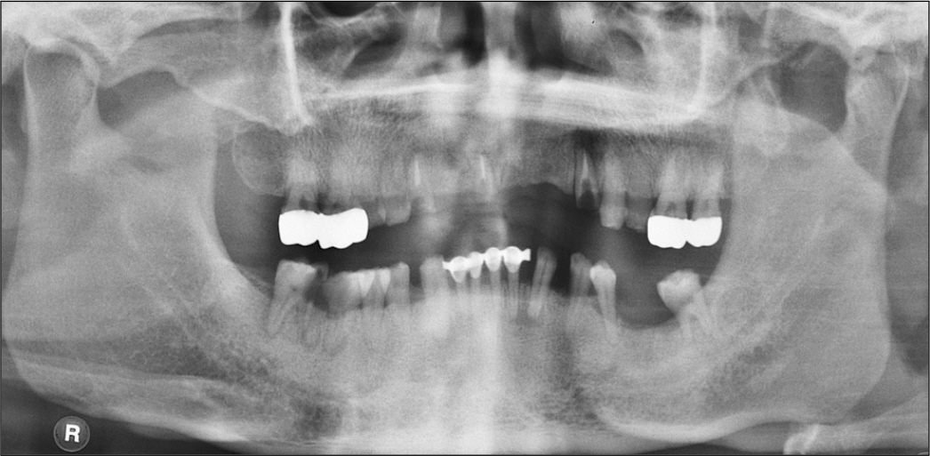
Fig. 2. Panoramic radiograph.
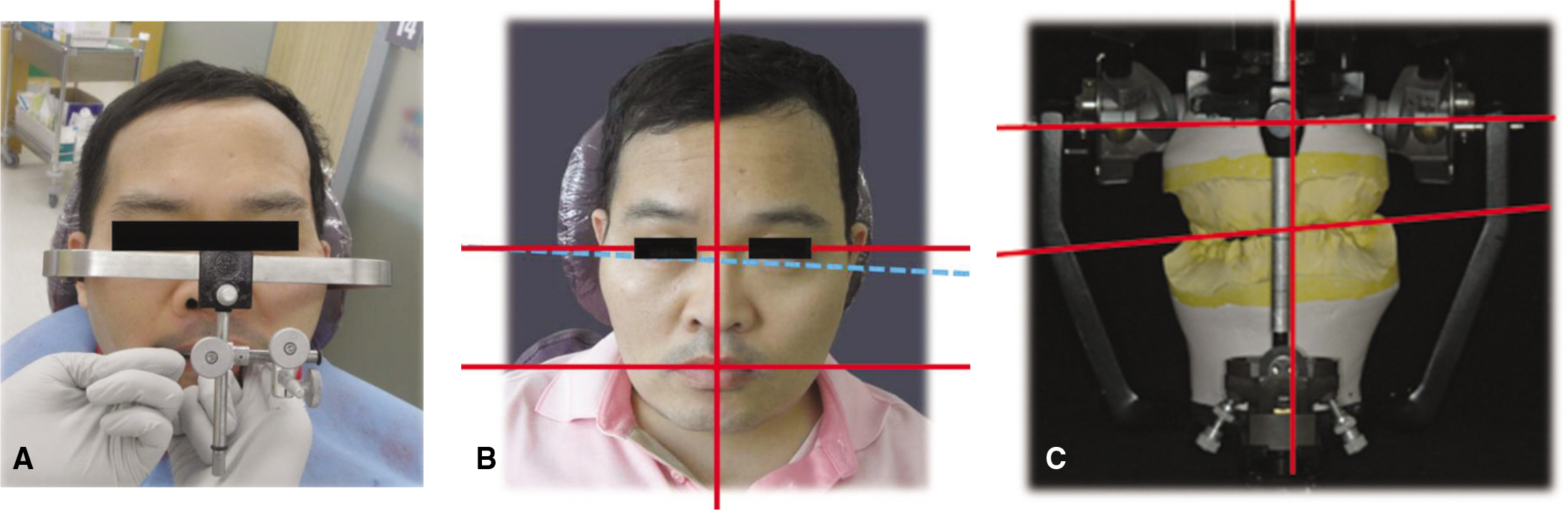
Fig. 3. Traditional ear-bow transfer. A: Traditional ear-bow transfer, B: Facial midline and occlusal plane, C: Articulator midline and occlusal plane.
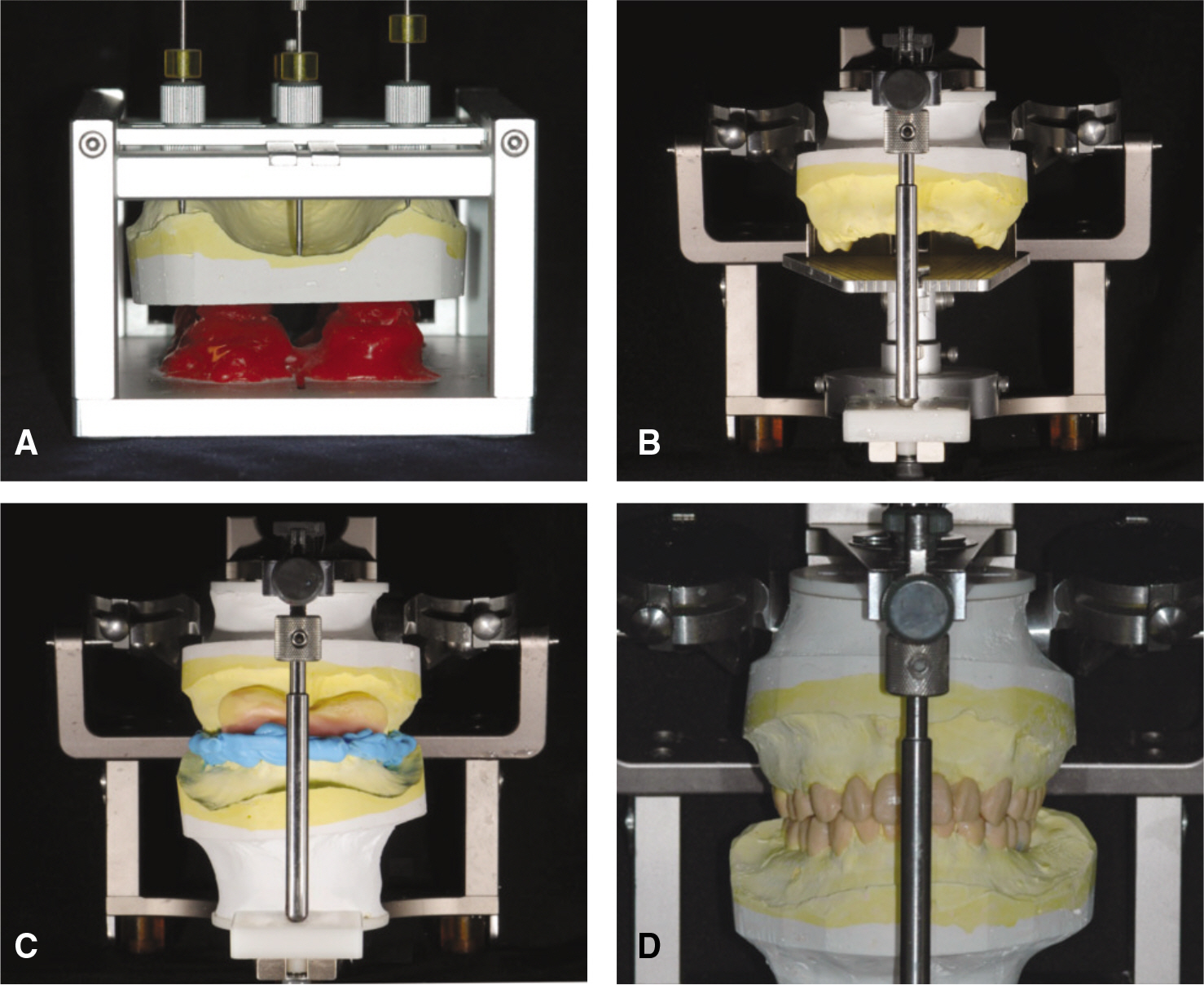
Fig. 4. Shilla system (Hamans, Tokyo, Japan). A: Shilla I - median-sagittal plane, B: Shilla II -occlusal plane determination, C: Gothic arch Tracer and mounting, D: Diagnostic wax-up.

Fig. 5. Temporary restoration. A: Intraoral photo, B: Extraoral photo.
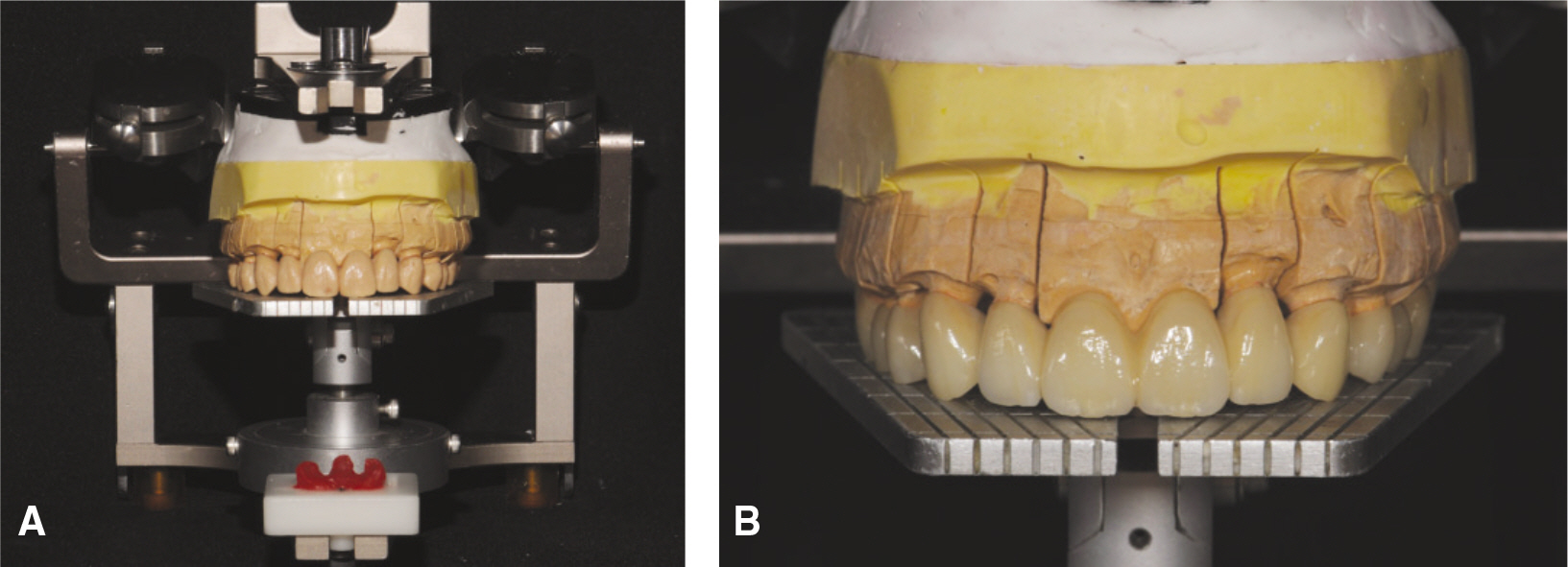
Fig. 6. Full contour wax-up. A: Full wax-up, B: Final maxillary restoration.

Fig. 7. Final restoration. A: Maxillary occlusal view, B: Mandibular occlusal view, C: Right buccal view, D: Left buccal view, E: Frontal view, F: Extraoral photograph.
Reference
-
1.Small BW. Occlusal plane analysis using the Broadrick flag. Gen Dent. 2005. 53:250–2.2.Choi DG., Bowley JF., Marx DB., Lee S. Reliability of an ear-bow arbitrary facebow transfer instrument. J Prosthet Dent. 1999. 82:150–6.
Article3.Turner KA., Missirlian DM. Restoration of the extremely worn dentition. J Prosthet Dent. 1984. 52:467–74.
Article4.Sato S., Hotta TH., Pedrazzi V. Removable occlusal overlay splint in the management of tooth wear: a clinical report. J Prosthet Dent. 2000. 83:392–5.
Article5.Hoyle DE. Fabrication of a customized anterior guide table. J Prosthet Dent. 1982. 48:490–1.
Article6.Toothaker RW., Graves AR. Custom adaptation of an occlusal plane analyzer to a semiadjustable articulator. J Prosthet Dent. 1999. 81:240–2.
Article7.Behrend DA. An esthetic control system for fixed and removable prosthodontics. J Prosthet Dent. 1985. 54:488–96.
Article8.Bowley JF., Michaels GC., Lai TW., Lin PP. Reliability of a facebow transfer procedure. J Prosthet Dent. 1992. 67:491–8.
Article9.Stawarczyk B., Ozcan M., Roos M., Trottmann A., Sailer I., Ha¨mmerle CH. Load-bearing capacity and failure types of anterior zirconia crowns veneered with overpressing and layering techniques. Dent Mater. 2011. 27:1045–53.
Article10.Wolfart S., Eschbach S., Scherrer S., Kern M. Clinical outcome of three-unit lithium-disilicate glass-ceramic fixed dental prostheses: up to 8 years results. Dent Mater. 2009. 25:e63–71.
Article11.Ishibe M., Raigrodski AJ., Flinn BD., Chung KH., Spiekerman C., Winter RR. Shear bond strengths of pressed and layered veneering ceramics to high-noble alloy and zirconia cores. J Prosthet Dent. 2011. 106:29–37.
Article
- Full Text Links
-
- Actions
-
Cited
- CITED
-
- Close
- Share
-
- Similar articles
-
- Full mounth rehabilitation using OP finder ® system for patient with inadequate occlusal plane and multiple occlusal wear tooth state: a case report
- Full mouth rehabilitation of class III patient with disharmonious occlusal plane: A case report
- Full-mouth rehabilitation in a patient with inclined occlusal plane and reduced vertical dimension by an attrition: A case report
- Full mouth rehabilitation through re-establishment of occlusal plane in partially edentulous patient with reduced vertical dimension accompanied by loss of posterior occlusal support
- Re-establishment of occlusal plane in a patient with a failed implant prosthesis
