Full-mouth rehabilitation in a patient with inclined occlusal plane and reduced vertical dimension by an attrition: A case report
- Affiliations
-
- 1Department of Prosthodontics, School of Dentistry, Chosun University, Gwangju, Republic of Korea. lkj1998@chosun.ac.kr
- KMID: 2444188
- DOI: http://doi.org/10.4047/jkap.2019.57.2.182
Abstract
- A proper vertical dimension and a harmonious occlusal plane are essential to satisfy a patient esthetically and functionally. A maxillomandibular occlusal vertical dimension is determined by the elevators which repeatedly contracts to a certain length, and a tooth location is determined by a maxillomandibular vertical dimension. The patient of this case came in with the incongruity of the lips and the occlusal plane. The result of clinical test showed the lack of length of the lower anterior due to the reduction of vertical dimension, the deep overbite of anterior, the excessive attrition of anterior, and the incongruity of occlusal plane. After the diagnostic wax-up, the temporary restoration was installed, and final prosthesis was installed after 6 months. As a result, the patient obtained a functionally and esthetically satisfying result.
MeSH Terms
Figure
-
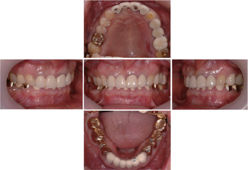
Fig. 1 Intraoral photograph before treatment. An inclined occlusal plane on the frontal view, a midline canting of maxillary central incisors, and attrition of PFM crowns were observed.

Fig. 2 Panoramic radiograph before the treatment.

Fig. 3 Facial photograph before the treatment. Remarkable inclined occlusal plane on the frontal view was observed. (I) Interpupillary line, (O) Frontal view of occlusal plane, (H) Horizontal plane which parallel to interpupillary line, (M) Midline which parallel to vertical line.
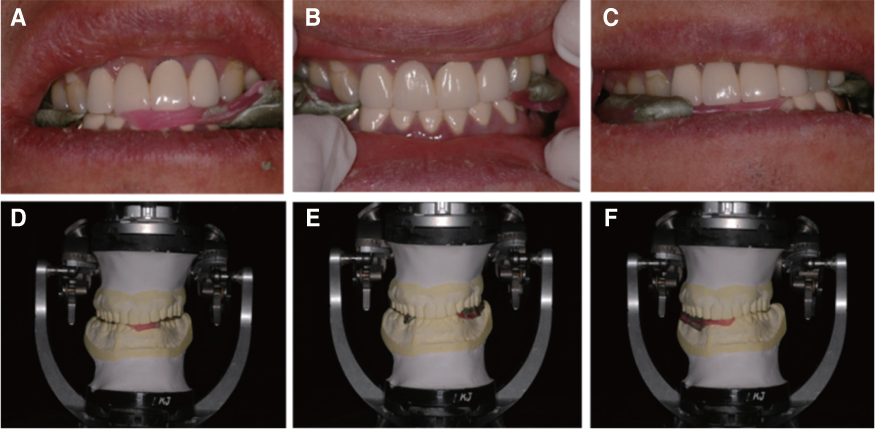
Fig. 4 Condylar guidance angle adjustment by check-bite method. (A, D) Right lateral movement, (B, E) Protrusive movement, (C, F) Left lateral movement.
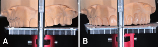
Fig. 5 Diagnostic wax-up model. (A) Diagnostic model, (B) Diagnostic wax-up.
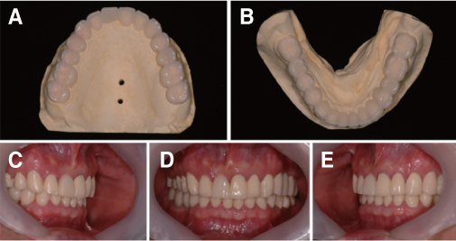
Fig. 6 Provisional restoration. (A) Upper, (B) Lower, (C) Right, (D) Frontal, (E) Left.
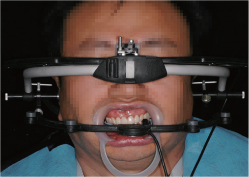
Fig. 7 Facial photograph with ARCUS Digma.

Fig. 8 Final prosthesis. (A) Right, (B) Frontal, (C) Left.

Fig. 9 Panoramic radiograph after treatment.

Fig. 10 Facial photograph after treatment. (I) Interpupillary line, (O) Frontal view of occlusal plane, (M) Midline which parallel to vertical line.
Reference
-
1. Mohindra NK, Bulman JS. The effect of increasing vertical dimension of occlusion on facial aesthetics. Br Dent J. 2002; 192:164–168.
Article2. The glossary of prosthodontic terms. 7th ed. J Prosthet Dent. 1999; 81:39–110.3. Pinho T, Neves M, Alves C. Multidisciplinary management including periodontics, orthodontics, implants, and prosthetics for an adult. Am J Orthod Dentofacial Orthop. 2012; 142:235–245.
Article4. Padwa BL, Kaiser MO, Kaban LB. Occlusal cant in the frontal plane as a reflection of facial asymmetry. J Oral Maxillofac Surg. 1997; 55:811–816.
Article5. Lussi A, Carvalho TS. Erosive tooth wear: a multifactorial condition of growing concern and increasing knowledge. Monogr Oral Sci. 2014; 25:1–15.
Article6. Loomans B, Opdam N, Attin T, Bartlett D, Edelhoff D, Frankenberger R, Benic G, Ramseyer S, Wetselaar P, Sterenborg B, Hickel R, Pallesen U, Mehta S, Banerji S, Lussi A, Wilson N. Severe tooth wear: European consensus statement on management guidelines. J Adhes Dent. 2017; 19:111–119.7. Muts EJ, van Pelt H, Edelhoff D, Krejci I, Cune M. Tooth wear: a systematic review of treatment options. J Prosthet Dent. 2014; 112:752–759.8. Mulay G, Dugal R, Buhranpurwala M. An evaluation of wear of human enamel opposed by ceramics of different surface finishes. J Indian Prosthodont Soc. 2015; 15:111–118.
Article9. Lee RL, Gregory GG. Gaining vertical dimension for the deep bite restorative patient. Dent Clin North Am. 1971; 15:743–763.10. Kim S, Han JS, Kim SH, Yoon HI, Yeo IS. Full mouth rehabilitation utilizing computer guided implant surgery and CAD/CAM. J Korean Acad Prosthodont. 2019; 57:57–65.
Article11. Tjan AH, Miller GD, The JG. Some esthetic factors in a smile. J Prosthet Dent. 1984; 51:24–28.
Article12. Turner KA, Missirlian DM. Restoration of the extremely worn dentition. J Prosthet Dent. 1984; 52:467–474.
Article13. Jo YJ, Jung S, Yang HS, Park SW, Lim HP, Yun KD, Park C. Full mouth rehabilitation of the patient with severe tooth loss and tooth wear with vertical dimension gaining: A case report. J Korean Acad Prosthodont. 2018; 56:302–307.
Article14. Koksal T, Dikbas I, Kazaoglu E. Alternative restorative approach for treatment of patient with extremely worn dentition. N Y State Dent J. 2009; 75:52–55.15. Abduo J, Lyons K. Clinical considerations for increasing occlusal vertical dimension: a review. Aust Dent J. 2012; 57:2–10.
Article16. Tallgren A, Lang BR, Walker GF, Ash MM Jr. Changes in jaw relations, hyoid position, and head posture in complete denture wearers. J Prosthet Dent. 1983; 50:148–156.
Article17. Dawson PE. Functional occlusion from: TMJ to smile design. St. Louis: Elsevier Health Science;2007. p. 430–452.18. Lerner J. A systematic approach to full-mouth reconstruction of the severely worn dentition. Pract Proced Aesthet Dent. 2008; 20:81–87.19. Rivera-Morales WC, Mohl ND. Relationship of occlusal vertical dimension to the health of the masticatory system. J Prosthet Dent. 1991; 65:547–553.
Article20. Braun S, Legan HL. Changes in occlusion related to the cant of the occlusal plane. Am J Orthod Dentofacial Orthop. 1997; 111:184–188.
Article
- Full Text Links
-
- Actions
-
Cited
- CITED
-
- Close
- Share
-
- Similar articles
-
- Full mouth rehabilitation of the elderly patient on anticoagulant medication with loss of vertical dimension due to severely worn dentition
- Full mouth rehabilitation using removable partial denture in patient with loss of vertical dimension due to worn dentition
- Full mouth rehabilitation through re-establishment of occlusal plane in partially edentulous patient with reduced vertical dimension accompanied by loss of posterior occlusal support
- Full mouth rehabilitation with extremely worn dentition
- Full mouth rehabilitation of the patient with severely worn dentition using monolithic zirconia prosthesis: A clinical report
