J Dent Rehabil Appl Sci.
2017 Sep;33(3):238-244. 10.14368/jdras.2017.33.3.238.
Full mouth rehabilitation with extremely worn dentition
- Affiliations
-
- 1Department of Prosthodontic Dentistry, Veterans Health Service Medical Center, Seoul, Republic of Korea. tchaski@daum.net
- KMID: 2394648
- DOI: http://doi.org/10.14368/jdras.2017.33.3.238
Abstract
- Pathologic attrition leads to pathologic damage on occlusal plane, functional disorders, occlusal disharmony, esthetic problems, pulpal lesion, temporomandibular joint (TMJ) disorder. In this case, treatment plan should be considered for possibility of vertical dimension loss, occlusal pattern, esthetics, phonetics, amount of vertical dimension increase. This case report was a 71-year-old man who had severely worn dentition. Full mouth rehabilitation was carried out with vertical dimension increase due to limited space for prosthesis. After evaluation of provisional restorations for patient's compliance, final restorations were fabricated and routine clinical assessments were made. This case presents that a satisfactory clinical result was achieved by restoring the worn dentition.
MeSH Terms
Figure
-
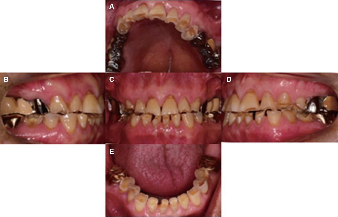
Fig. 1 Intraoral photograph before treatment. (A) Upper view, (B) Right view, (C) Frontal view, (D) Left view, (E) Lower view.
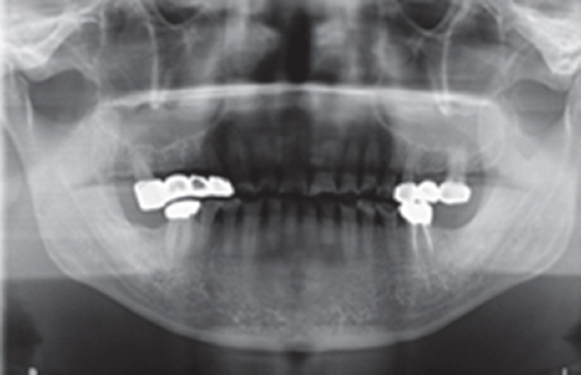
Fig. 2 Panoramic radiograph before treatment.
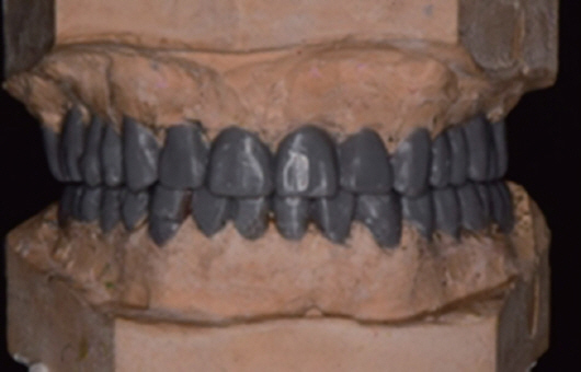
Fig. 3 Diagnostic waxup cast model.
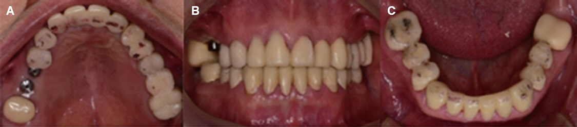
Fig. 4 Provisional restoration. (A) Upper view, (B) Frontal view, (C) Lower view.
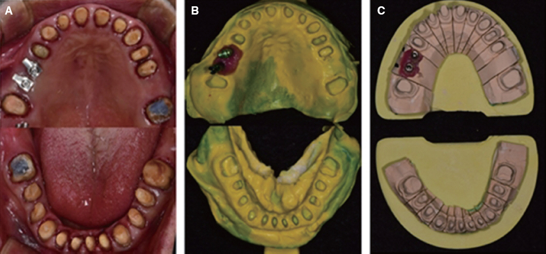
Fig. 5 (A) Tooth preparation, (B) Final impression taking, (C) Die preparation.
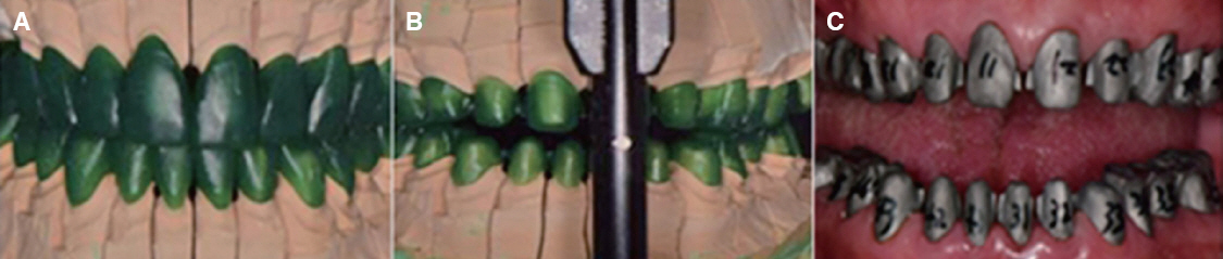
Fig. 6 (A) Full contour wax up, (B) Cut back, (C) Metal coping try in.
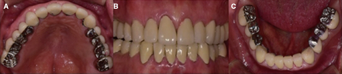
Fig. 7 Definitive prosthesis. (A) Upper view, (B) Frontal view, (C) Lower view.
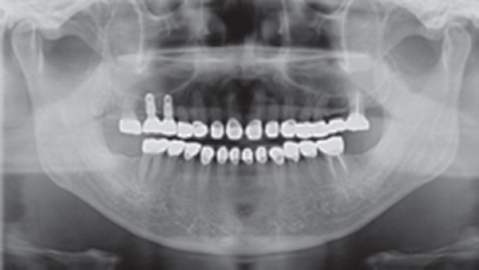
Fig. 8 Panoramic radiograph after treatme
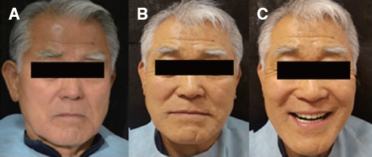
Fig. 9 Extraoral photograph. (A) Before treatment, frontal view, (B) After treatment, frontal view, (C) After treatment, smile.
Reference
-
References
1. Mulay G, Dugal R, Buhranpurwala M. An evaluation of wear of human enamel opposed by ceramics of different surface finishes. J Indian Prosthodont Soc. 2015; 15:111–8. DOI: 10.4103/0972-4052.155031. PMID: 26929496. PMCID: PMC4762315.2. Ibbetson RJ, Setchell DJ. Treatment of the worn dentition:2. Dent Update. 1989; 16:300–2. 305-7. PMID: 2638262.3. Silverman MM. The speaking method in measuring vertical dimension 1952. J Prosthet Dent. 2001; 85:427–31. DOI: 10.1067/mpr.2001.116139. PMID: 11357066.4. Silverman MM. Determination of vertical dimension by phonetics. J Prosthet Dent. 1956; 6:467–71. DOI: 10.1016/0022-3913(56)90091-9.5. Turner KA, Missirlian DM. Restoration of the extremely worn dentition. J Prosthet Dent. 1984; 52:467–74. DOI: 10.1016/0022-3913(84)90326-3.6. Johnson A, Wildgoose DG, Wood DJ. The determination of freeway space using two different methods. J Oral Rehabil. 2002; 29:1010–3. DOI: 10.1046/j.1365-2842.2002.00950.x.7. Park JH, Jeong CM, Jeon YC, Lim JS. A study on the occlusal plane and the vertical dimension in Korean adults with natural dentition. J Korean Acad Prosthodont. 2005; 43:41–51.8. Willis FM. Features of the face involved in full denture prosthesis. Dent Cosmos. 1935; 77:851–4.9. Dawson PE. Functional occlusion: from TMJ to smile design. St. Louis: Elsevier Health Sciences;2007. p. 430–52.10. Rivera-Morales WC, Mohl ND. Relationship of occlusal vertical dimension to the health of the masticatory system. J Prosthet Dent. 1991; 65:547–53. DOI: 10.1016/0022-3913(91)90298-B.11. Abduo J, Lyons K. Clinical considerations for increasing occlusal vertical dimension: a review. Aust Dent J. 2012; 57:2–10. DOI: 10.1111/j.1834-7819.2011.01640.x. PMID: 22369551.12. Oh YR, Lee SB, Park NS, Choi DG. A study of intraoral anatomic landmarks of Korean adult-upper jaw. J Korean Acad Prosthodont. 1995; 33:753–68.
- Full Text Links
-
- Actions
-
Cited
- CITED
-
- Close
- Share
-
- Similar articles
-
- Complete mouth rehabilitation with vertical dimension increase in patient with extremely worn dentition
- Full-mouth rehabilitation without changing the vertical dimension in patient with worn dentition
- Full mouth rehabilitation using zirconia crown in severe worn dentition: a case report
- Mandibular full arch rehabilitation of a patient with severely worn dentition after completion of maxillary full arch rehabilitation
- Full mouth rehabilitation of a patient with worn dentition and loss of posterior support by vertical dimension reestablishment: a clinical report
