Image Findings of Primary Squamous Cell Carcinoma of the Pancreas in Patient with Chronic Pancreatitis: A Case Report
- Affiliations
-
- 1Department of Radiology, Inha University School of Medicine, Inha University Hospital, Korea. mykim@inha.ac.kr
- 2Department of Surgery, Inha University School of Medicine, Inha University Hospital, Korea.
- 3Department of Pathology, Inha University School of Medicine, Inha University Hospital, Korea.
- 4Biology, New York University, College of Arts and Science, New York University, Korea.
- KMID: 2000065
- DOI: http://doi.org/10.13104/jksmrm.2011.15.2.160
Abstract
- Squamous cell carcinoma of the pancreas is a rare, uncommon tumor that is characterized by squamous metaplasia of the ductal columnar cells. We report the image findings of a rare case of the pancreatic squamous cell carcinoma associated with chronic pancreatitis.
Keyword
Figure
-
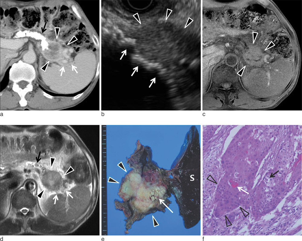
Fig. 1 (a) Contrast enhanced CT scan shows heterogeneously peripheral enhancing solid mass (arrowheads) and perilesional invasion (white arrows) on equilibrium phase. Note dense calcifications (black arrow) within diffuse atrophic change of pancreas, suggesting chronic pancreatitis. (b) EUS (endoscopic ultrasonography) shows lobulating solid mass (arrowheads) in tail portion of pancreas, and multiple hyperechogeic stones (arrows) in tail of pancreas. (c) Axial T1-weighted MR image shows iso-signal intensity of the pancreatic mass lesion (arrow heads) than that of spleen. (d) Axial T2-weighted MR image shows heterogeneously hyper intense mass (arrow heads) in pancreatic tail and perilesional infiltration in splenic hilum (white arrows). Signal intensity of mass lesion is higher than that of spleen. Intraductal stone (black arrow) and parenchyma atrophy of pancreas are suggestive findings of chronic pancreatitis. (e) Surgical specimen shows solid mass (arrowheads) in distal pancreas extends to the peripancreatic soft tissue and hilum of spleen (S). Note the small intraductal and stones (arrow). (f) Histologic section demonstrating a squamous cell carcinoma with intraparenchymal tumor nest (arrow heads), keratin pearl (white arrow), and clear cell change (black arrow) (H & E stain, 200 × magnification).
Cited by 1 articles
-
Squamous Cell Carcinoma of the Pancreas: A Case Report
Se Hoon Park, Ju Wan Choi, Ji Yeol Shin, Seong Hoon Kim
J Korean Soc Radiol. 2018;79(4):191-195. doi: 10.3348/jksr.2018.79.4.191.
Reference
-
1. Al-Sherhri A, Silverman S, King KM. Squamous cell carcinoma of the pancreas. Current Oncology. 2008. 15:293–297.2. Lowry CC, Whitaker HW Jr, Greiner DJ. Squamous cell carcinoma of the pancreas. South Med J. 1949. 42:753–757.3. Mulkeen AL, Yoo PS, Cha C. Less common neoplasms of the pancreas. World J Gastroenterol. 2006. 12(20):3180–3185.4. Brown HA, Dotto J, Robert M, Salem RR. Squamous cell carcinoma of the pancreas. J Clin Gastroenterol. 2005. 39:915–919.5. Anagnostopoulos GK, Aithal GP, Ragunath K, Kaye P, Rowlands BJ. Squamous cell carcinoma of the pancreas: report of a case and review of the literature. JOP. 2006. 7:47–50.6. Ishikawa O, Matsui Y, Aoki I, Iwanaga T, Terasawa T, Wada A. Adenosquamous carcinoma of the pancreas: a clinicopathologic study and report of three cases. Cancer. 1980. 46:1192–1196.7. Nakashima H, Hayakawa T, Hoshino M, et al. Squamous cell carcinoma of the pancreas with massive invasion of the retroperitoneum. Intern Med. 1995. 34:61–64.8. Fajardo LL, Yoshino MT, Chernin MM. Computed tomography findings in squamous cell carcinoma of the pancreas. J Comput Assist Tomogr. 1988. 12:138–139.9. Sprayregen S, Schoenbaum SW, Messinger NH. Angiographic features of squamous cell carcinoma of the pancreas. J Can Assoc Radiol. 1975. 26:122–124.10. Minami T, Fukui K, Morita Y, et al. A case of squamous cell carcinoma of the pancreas with an initial symptom of tarry stool. J Gastroenterol Hepatol. 2001. 16:1077–1079.11. Yu MH, Lee JY, Kim MA, et al. MR imaging features of small solid pseudopapillary tumors: retrospective differentiation from other small solid pancreatic tumors. AJR Am J Roentgenol. 2010. 195:1324–1332.12. Paciorek ML, Ross GJ. MR imaging of primary pancreatic leiomyosarcoma. Br J Radiol. 1998. 71:561–563.13. Brayko CM, Doll DC. Squamous cell carcinoma of the pancreas associated with hypercalcemia. Gastroenterology. 1982. 83:1297–1299.
- Full Text Links
-
- Actions
-
Cited
- CITED
-
- Close
- Share
-
- Similar articles
-
- Squamous Cell Carcinoma of the Pancreas: A Case Report
- Squamous cell carcinoma of the pancreas with a pancreatic intraductal papillary mucinous neoplasm: a case report
- Primary Squamous Cell Carcinoma of the Ureter after Reterocutaneoustomy
- Duodenal ectopic pancreas complicated by chronic pancreatitis and pseudocyst formation: a case report
- CT Findings of Primary Squamous Cell Carcinoma of the Stomach: A Case Report
