Squamous cell carcinoma of the pancreas with a pancreatic intraductal papillary mucinous neoplasm: a case report
- Affiliations
-
- 1Department of Radiology, Biomedical Research Institute, Pusan National University Hospital, Pusan National University School of Medicine, Busan, Korea
- KMID: 2556801
- DOI: http://doi.org/10.7180/kmj.23.123
Abstract
- Squamous cell carcinoma (SCC) of the pancreas is very rare. No reports have described SCC accompanied by intraductal papillary mucinous neoplasm (IPMN) of the pancreas. This report presents the first known case of SCC with IPMN of the pancreas in a 71-year-old man, with a focus on radiologic findings. Here, the imaging features of SCC with IPMN of the pancreas were similar to those of IPMN of the pancreas with high-risk stigmata features.
Keyword
Figure
-
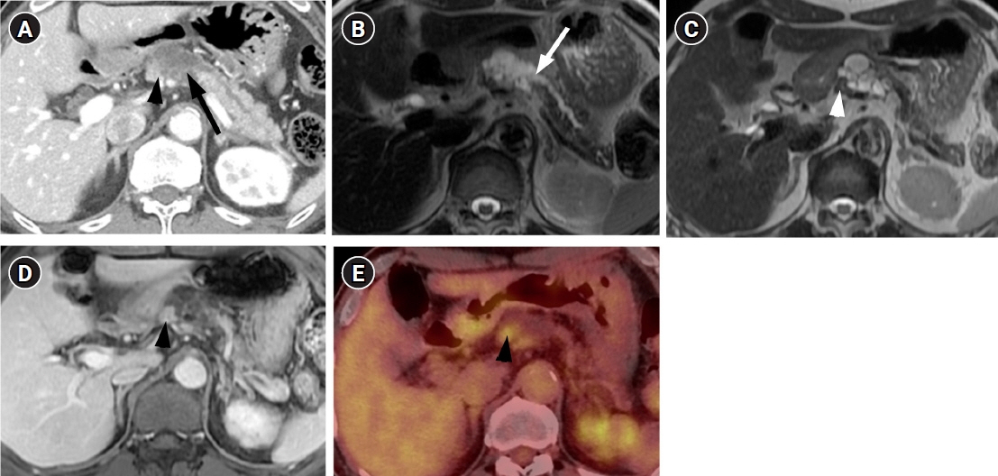
Fig. 1. CT, MRI, and PET-CT findings of squamous cell carcinoma with pancreatic intraductal papillary mucinous neoplasm in a 71-year-old man. (A) Contrast-enhanced CT shows a multiseptated cystic lesion (arrow) with an enhancing mural nodule (arrowhead) in the body of the pancreas. (B, C) Axial T2-weighted MRI shows a communication (arrow in B) between the pancreatic cystic lesion and main pancreatic duct. A T2 isointense mural nodule (arrowhead in C) is also noted in the cystic lesion. (D) Axial contrast-enhanced T1-weighted MRI shows the enhancing mural nodule (arrowhead) in the cystic lesion. (E) PET- CT shows FDG uptake of the mural nodule (arrowhead) in the cystic lesion. CT, computed tomography; MRI, magnetic resonance imaging; PET-CT, positron emission tomography-CT.
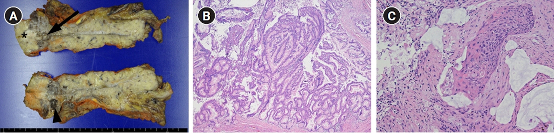
Fig. 2. Histopathologic findings of squamous cell carcinoma with pancreatic intraductal papillary mucinous neoplasm in a 71-year-old man. (A) The cut section of the gross specimen shows a macrocystic change (arrowhead) in the pancreas and dilatated main pancreatic duct (arrow). There is a relatively well-defined intramural solid nodule (asterisk) on the proximal side of the pancreatic duct. (B) Low-power magnification shows the dilated pancreatic duct lined by gastric type epithelial cells with low- to high-grade dysplasia and forming papillary structures, which consists with intraductal papillary mucinous neoplasm of the pancreas (hematoxylin and eosin [H&E] stain, ×40). (C) High-power magnification shows that the mural nodule demonstrates keratinized squamous epithelial cells with intercellular bridging and high-grade nuclear atypia, which are compatible with squamous cell carcinoma (H&E stain, ×200).
Reference
-
References
1. Castellano-Megias VM, Andres CI, Lopez-Alonso G, Colina-Ruizdelgado F. Pathological features and diagnosis of intraductal papillary mucinous neoplasm of the pancreas. World J Gastrointest Oncol. 2014; 6:311–24.2. Tanaka M, Fernandez-Del Castillo C, Kamisawa T, Jang JY, Levy P, Ohtsuka T, et al. Revisions of international consensus Fukuoka guidelines for the management of IPMN of the pancreas. Pancreatology. 2017; 17:738–53.3. Takanami K, Hiraide T, Tsuda M, Nakamura Y, Kaneta T, Takase K, et al. Additional value of FDG PET/CT to contrast-enhanced CT in the differentiation between benign and malignant intraductal papillary mucinous neoplasms of the pancreas with mural nodules. Ann Nucl Med. 2011; 25:501–10.44. Kim JH, Eun HW, Kim KW, Lee JY, Lee JM, Han JK, et al. Intraductal papillary mucinous neoplasms with associated invasive carcinoma of the pancreas: imaging findings and diagnostic performance of MDCT for prediction of prognostic factors. AJR Am J Roentgenol. 2013; 201:565–72.5. Okamura Y, Sugimoto H, Fujii T, Nomoto S, Takeda S, Nakao A. Adenosquamous carcinoma arising in an intraductal papillary mucinous neoplasm of the pancreas. Pancreas. 2010; 39:945–7.6. Makarova-Rusher OV, Ulahannan S, Greten TF, Duffy A. Pancreatic squamous cell carcinoma: a population-based study of epidemiology, clinicopathologic characteristics and outcomes. Pancreas. 2016; 45:1432–7.7. Ntanasis-Stathopoulos I, Tsilimigras DI, Georgiadou D, Kanavidis P, Riccioni O, Salla C, et al. Squamous cell carcinoma of the pancreas: a systematic review and pooled survival analysis. Eur J Cancer. 2017; 79:193–204.8. Fajardo LL, Yoshino MT, Chernin MM. Computed tomography findings in squamous cell carcinoma of the pancreas. J Comput Tomogr. 1988; 12:138–9.9. Kim JB, Kim MY, Suh CH, Lee KY, Joo YC, Cho JY. Image findings of primary squamous cell carcinoma of the pancreas in patient with chronic pancreatitis: a case report. J Korean Soc Magn Reson Med. 2011; 15:160–4.10. Lu T, Li X, Zhou Y. Pancreatic metastasis from squamous cell lung cancer: computed tomography and magnetic resonance imaging findings. J Int Med Res. 2021; 49:300060521996188.
- Full Text Links
-
- Actions
-
Cited
- CITED
-
- Close
- Share
-
- Similar articles
-
- Malignant Pancreatic Intraductal Papillary Mucinous Neoplasm with Splenic Invasion: A Case Report
- A Case of Intraductal Papillary Mucinous Neoplasm Arising from Santorini's Duct in a Patient with Complete Type of Pancreas Divisum
- Intraductal Papillary Mucinous Tumor Simultaneously Involving the Liver and Pancreas: A Case Report
- A case of intraductal papillary tumor of pancreas associated with mucinous ductal ectasia
- Comparison of Mucinous Cystic Tumor and Intraductal Papillary Mucinous Tumor
