Evaluation of the palatal soft tissue thickness by cone-beam computed tomography
- Affiliations
-
- 1Department of Orthodontics, Seoul St. Mary's Hospital, The Catholic University of Korea, Seoul, Korea.
- 2Division of Orthodontics, Department of Dentistry, St. Vincent's Hospital, The Catholic University of Korea, Suwon, Korea. seonghh@hotmail.com
- KMID: 1976681
- DOI: http://doi.org/10.4041/kjod.2012.42.6.291
Abstract
OBJECTIVE
The purposes of this study were to measure the palatal soft tissue thickness at popular placement sites of temporary anchorage devices (TADs) by cone-beam computed tomography (CBCT) and evaluate the age, gender, and positional differences in this parameter.
METHODS
The study sample consisted of 23 children (10 boys and 13 girls; mean age, 10.87 +/- 1.24 years; range, 6.7 to 12.6 years) and 27 adults (14 men and 13 women; mean age, 21.35 +/- 1.14 years; range, 20.0 to 23.8 years). Nine mediolateral and nine anteroposterior intersecting reference lines were drawn on CBCT scans of the 50 subjects, and the resultant measurement areas were designated according to their mediolateral (i.e., lateral, medial, and sutural) and anteroposterior (i.e., anterior, middle, and posterior) positions. Repeated-measures analysis of variance was performed to analyze intragroup and intergroup differences.
RESULTS
No significant age and gender differences were found (p = 0.309 and 0.124, respectively). Further, no significant anteroposterior change was observed (p = 0.350). However, the lateral area presented the thickest soft tissue whereas the sutural area had the thinnest soft tissue (p < 0.001).
CONCLUSIONS
Clinical selection of the placement sites of TADs should be guided by knowledge of the positional variations in the palatal soft tissue thickness in addition to other contributing factors of TAD stability.
Keyword
Figure
-

Figure 1 Anteroposterior (AP) and mediolateral (ML) reference lines forming 81 intersection points for measuring the palatal soft tissue thickness.
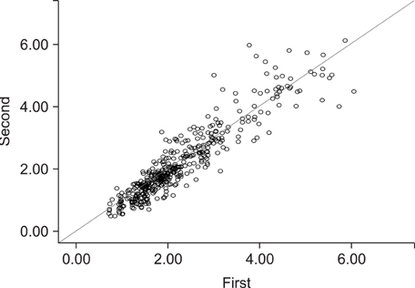
Figure 2 Bland-Altman plot of the intra-examiner assessment reliability (unit: mm).

Figure 3 Changes in the palatal soft tissue thickness according to the mediolateral positions.

Figure 4 Soft tissue thickness at different mediolateral positions of the palate.
Cited by 3 articles
-
Comparison of treatment effects between the modified C-palatal plate and cervical pull headgear for total arch distalization in adults
Chong Ook Park, Noor Laith Sa'aed, Mohamed Bayome, Jae Hyun Park, Yoon-Ah Kook, Young-Seok Park, Seong Ho Han
Korean J Orthod. 2017;47(6):375-383. doi: 10.4041/kjod.2017.47.6.375.Distalization with a modified C-palatal plate for severe upper crowding and a missing lower incisor
Jae Hyun Park, Traci Saito, Sun Kyong Yoo, Mohammed Alfaifi, Yoon-Ah Kook
Korean J Orthod. 2020;50(1):52-62. doi: 10.4041/kjod.2020.50.1.52.Displacement and stress distribution of the maxillofacial complex during maxillary protraction using palatal plates: A three-dimensional finite element analysis
Jusuk Eom, Mohamed Bayome, Jae Hyun Park, Hee Jin Lim, Yoon-Ah Kook, Seong Ho Han
Korean J Orthod. 2018;48(5):304-315. doi: 10.4041/kjod.2018.48.5.304.
Reference
-
1. Kinzinger GS, Eren M, Diedrich PR. Treatment effects of intraoral appliances with conventional anchorage designs for non-compliance maxillary molar distalization: a literature review. Eur J Orthod. 2008. 30:558–571.
Article2. Papadopoulos MA, Tarawneh F. The use of miniscrew implants for temporary skeletal anchorage in orthodontics: a comprehensive review. Oral Surg Oral Med Oral Pathol Oral Radiol Endod. 2007. 103:e6–e15.
Article3. Hoste S, Vercruyssen M, Quirynen M, Willems G. Risk factors and indications of orthodontic temporary anchorage devices: a literature review. Aust Orthod J. 2008. 24:140–148.4. Reynders R, Ronchi L, Bipat S. Mini-implants in orthodontics: a systematic review of the literature. Am J Orthod Dentofacial Orthop. 2009. 135:564.e1–564.e19.
Article5. Chen YJ, Chang HH, Huang CY, Hung HC, Lai EH, Yao CC. A retrospective analysis of the failure rate of three different orthodontic skeletal anchorage systems. Clin Oral Implants Res. 2007. 18:768–775.
Article6. Kyung SH, Lee JY, Shin JW, Hong C, Dietz V, Gianelly AA. Distalization of the entire maxillary arch in an adult. Am J Orthod Dentofacial Orthop. 2009. 135:4 Suppl. S123–S132.
Article7. Kook YA, Kim SH, Chung KR. A modified palatal anchorage plate for simple and efficient distalization. J Clin Orthod. 2010. 44:719–730.8. Sandler J, Benson PE, Doyle P, Majumder A, O'Dwyer J, Speight P, et al. Palatal implants are a good alternative to headgear: a randomized trial. Am J Orthod Dentofacial Orthop. 2008. 133:51–57.
Article9. Greenberg J, Laster L, Listgarten MA. Transgingival probing as a potential estimator of alveolar bone level. J Periodontol. 1976. 47:514–517.
Article10. Wara-aswapati N, Pitiphat W, Chandrapho N, Rattanayatikul C, Karimbux N. Thickness of palatal masticatory mucosa associated with age. J Periodontol. 2001. 72:1407–1412.
Article11. Eger T, Müller HP, Heinecke A. Ultrasonic determination of gingival thickness: subject variation and influence of tooth type and clinical features. J Clin Periodontol. 1996. 23:839–845.
Article12. Müller HP, Schaller N, Eger T, Heinecke A. Thickness of masticatory mucosa. J Clin Periodontol. 2000. 27:431–436.
Article13. Song JE, Um YJ, Kim CS, Choi SH, Cho KS, Kim CK, et al. Thickness of posterior palatal masticatory mucosa: the use of computerized tomography. J Periodontol. 2008. 79:406–412.
Article14. Ueno D, Sato J, Igarashi C, Ikeda S, Morita M, Shimoda S, et al. Accuracy of oral mucosal thickness measurements using spiral computed tomography. J Periodontol. 2011. 82:829–836.
Article15. Januário AL, Barriviera M, Duarte WR. Soft tissue cone-beam computed tomography: a novel method for the measurement of gingival tissue and the dimensions of the dentogingival unit. J Esthet Restor Dent. 2008. 20:366–373.
Article16. Barriviera M, Duarte WR, Januário AL, Faber J, Bezerra AC. A new method to assess and measure palatal masticatory mucosa by cone-beam computerized tomography. J Clin Periodontol. 2009. 36:564–568.
Article17. Fu JH, Yeh CY, Chan HL, Tatarakis N, Leong DJ, Wang HL. Tissue biotype and its relation to the underlying bone morphology. J Periodontol. 2010. 81:569–574.
Article18. Kim HJ, Yun HS, Park HD, Kim DH, Park YC. Soft-tissue and cortical-bone thickness at orthodontic implant sites. Am J Orthod Dentofacial Orthop. 2006. 130:177–182.
Article19. Moon SH, Park SH, Lim WH, Chun YS. Palatal bone density in adult subjects: implications for mini-implant placement. Angle Orthod. 2010. 80:137–144.
Article20. Guerrero ME, Jacobs R, Loubele M, Schutyser F, Suetens P, van Steenberghe D. State-of-the-art on cone beam CT imaging for preoperative planning of implant placement. Clin Oral Investig. 2006. 10:1–7.
Article21. Cha BK, Lee YH, Lee NK, Choi DS, Baek SH. Soft tissue thickness for placement of an orthodontic miniscrew using an ultrasonic device. Angle Orthod. 2008. 78:403–408.
Article
- Full Text Links
-
- Actions
-
Cited
- CITED
-
- Close
- Share
-
- Similar articles
-
- Detection of maxillary second molar with two palatal roots using cone beam computed tomography: a case report
- Quantitative evaluation of palatal bone thickness in patients with normal and open vertical skeletal configurations using cone-beam computed tomography
- Quantitative cone-beam computed tomography evaluation of hard and soft tissue thicknesses in the midpalatal suture region to facilitate orthodontic mini-implant placement
- Assessment of the relationship between the maxillary molars and adjacent structures using cone beam computed tomography
- Three-dimensional imaging modalities in endodontics
