J Adv Prosthodont.
2015 Feb;7(1):62-68. 10.4047/jap.2015.7.1.62.
Effects of counter torque and transposition (transfer) of installed implants timing on their integration in dog tibia
- Affiliations
-
- 1Department of Periodontics, Dental Branch, Islamic Azad University, Tehran, Iran. shfathi7@gmail.com
- 2Postgraduate of Oral Medicine, Dental Branch, Hamadan University of Medical Sciences, Hamadan, Iran.
- 3Department of Periodontics, Dental Research Center, Dental School, Shahid Beheshti University of Medical Sciences, Tehran, Iran.
- KMID: 1974893
- DOI: http://doi.org/10.4047/jap.2015.7.1.62
Abstract
- PURPOSE
The purpose of this research was to evaluate the amount of reosseointegration after counter torquing (reverse torque) and transposing the installed implants at different times.
MATERIALS AND METHODS
This study was done on ten tibiae of five cross-bred dogs. At the first day one implant was installed in each tibia. After one week half of the implants were randomly counter torqued (1WCT) and the other half were explanted and reimplanted in a new juxtaposition site (transposed)(1WT). At the same time three new implants were installed in each dog, one of them was considered as one week control (1WC) and remaining two as 8 week groups (8WCT&8WT). After eight weeks the 1WCT and 1WT implants were loosened by counter torque and the quantity of needed force for liberation was measured with the digital device (BGI). At the same time one implant was installed in each dog as eight week control (8WC) and the same protocol was repeated for 8 week groups after another 8 weeks.
RESULTS
All implants were osseointegrated. Mean quantities of osseointegration in case groups indicated better amounts rather than control groups.
CONCLUSION
Counter torque or transposition of the installed implants one week or eight weeks after the implantation did lead to osseointegration.
Keyword
MeSH Terms
Figure
-
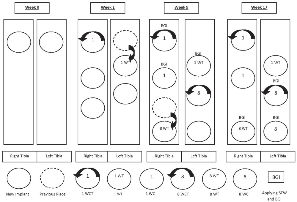
Fig. 1 Timeline graphic of implants.
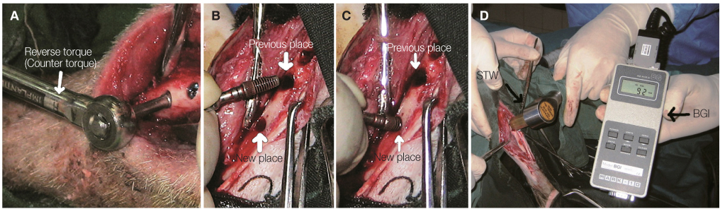
Fig. 2 Overview of surgical procedures in this study. (A) Performing counter torque the implant, (B) Exiting the implant of own cavity, (C) Transposing the implant to new cavity near the previous place immediately, (D) Reflecting periosteal flap at 17th week, (E) Applying STW and BGI for recording the peak force required to detach the implant.4
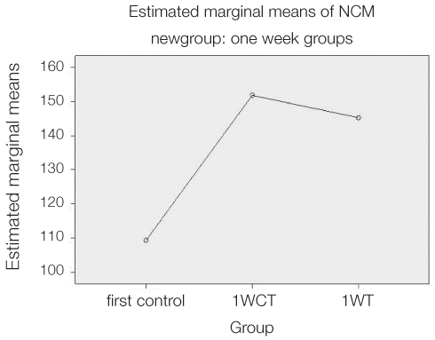
Fig. 3 Counter torque value (Ncm) between 3 experimental groups (first control - 1 WCT - 1 WT).
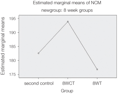
Fig. 4 Counter torque value (Ncm) between 3 experimental groups (second control - 8 WCT - 8 WT).
Reference
-
1. Ivanoff CJ, Sennerby L, Lekholm U. Reintegration of mobilized titanium implants. An experimental study in rabbit tibia. Int J Oral Maxillofac Surg. 1997; 26:310–315.2. Moriya K, Maruo Y, Minagi S. Does rotational strain at screw tightening affect the attainment or maintenance of osseointegration? Clin Oral Implants Res. 2006; 17:451–458.3. Lang LA, May KB, Wang RF. The effect of the use of a counter-torque device on the abutment-implant complex. J Prosthet Dent. 1999; 81:411–417.4. Lucente J, Galante J, Trisi P, Kenealy JN. Reintegration success of osseotite implants after intentional countertorque liberation in the endentulous human mandible. Implant Dent. 2006; 15:178–185.5. Albrektsson T, Zarb G, Worthington P, Eriksson AR. The long-term efficacy of currently used dental implants: a review and proposed criteria of success. Int J Oral Maxillofac Implants. 1986; 1:11–25.6. Smith DE, Zarb GA. Criteria for success of osseointegrated endosseous implants. J Prosthet Dent. 1989; 62:567–572.7. Misch CE, Perel ML, Wang HL, Sammartino G, Galindo-Moreno P, Trisi P, Steigmann M, Rebaudi A, Palti A, Pikos MA, Schwartz-Arad D, Choukroun J, Gutierrez-Perez JL, Marenzi G, Valavanis DK. Implant success, survival, and failure: the International Congress of Oral Implantologists (ICOI) Pisa Consensus Conference. Implant Dent. 2008; 17:5–15.8. Sakka S, Coulthard P. Implant failure: etiology and complications. Med Oral Patol Oral Cir Bucal. 2011; 16:e42–e44.9. Lekholm U, Adell R, Brånemark PI. Complications. In : Brånemark PI, Zarb GA, Albrektsson T, editors. Tissue-integrated prostheses: Osseointegration in clinical dentistry. Chicago: Quintessence;1985. p. 233–240.10. Sennerby L, Meredith N. Implant stability measurements using resonance frequency analysis: biological and biomechanical aspects and clinical implications. Periodontol 2000. 2008; 47:51–66.11. Renvert S, Polyzois I, Maguire R. Re-osseointegration on previously contaminated surfaces: a systematic review. Clin Oral Implants Res. 2009; 20:216–227.12. Friberg B, Jemt T, Lekholm U. Early failures in 4,641 consecutively placed Brånemark dental implants: a study from stage 1 surgery to the connection of completed prostheses. Int J Oral Maxillofac Implants. 1991; 6:142–146.13. Sullivan DY, Sherwood RL, Collins TA, Krogh PH. The reverse-torque test: a clinical report. Int J Oral Maxillofac Implants. 1996; 11:179–185.14. Turner AS, Mcllwraith CW, Hull BL. Techniques in large animal surgery. 2nd ed. Philadelphia: Lea & Febiger;1989. p. 87–89.15. Carvalho CM, Carvalho LF, Costa LJ, Sa MJ, Figueiredo CR, Azevedo AS. Titanium implants: a removal torque study in osteopenic rabbits. Indian J Dent Res. 2010; 21:349–352.16. Davies JE. Mechanisms of endosseous integration. Int J Prosthodont. 1998; 11:391–401.17. Morberg P, Albrektsson T. A histomorphometric and removal torque analysis of c.p. titanium implants inserted in reamed bone beds with and without acrylic cement. J Mater Sci Mater Med. 1992; 3:170–174.18. Roccuzzo M, Bunino M, Prioglio F, Bianchi SD. Early loading of sandblasted and acid-etched (SLA) implants: a prospective split-mouth comparative study. Clin Oral Implants Res. 2001; 12:572–578.19. Cochran DL, Buser D, ten Bruggenkate CM, Weingart D, Taylor TM, Bernard JP, Peters F, Simpson JP. The use of reduced healing times on ITI implants with a sandblasted and acid-etched (SLA) surface: early results from clinical trials on ITI SLA implants. Clin Oral Implants Res. 2002; 13:144–153.20. Salvi GE, Gallini G, Lang NP. Early loading (2 or 6 weeks) of sandblasted and acid-etched (SLA) ITI implants in the posterior mandible. A 1-year randomized controlled clinical trial. Clin Oral Implants Res. 2004; 15:142–149.21. Poggio CE, Salvato A. Implant repositioning for esthetic reasons: a clinical report. J Prosthet Dent. 2001; 86:126–129.22. Lindhe J, Lang NP, Karring T. Clinical periodontology and implant dentistry. 4th ed. China: Blackwell;2003. p. 866–874.23. Misch CE. Dental Implant prosthodontics Misch. China: Mosby Elsevier;2005. p. 340.24. Frost HM. The regional acceleratory phenomenon. Intermediary organization of the skeleton. Boca Raton, FL, USA: CRC Press;1986. p. 109–129.25. Lundgren D, Lundgren AK, Sennerby L. The effect of mechanical intervention on jaw bone density. Clin Oral Implants Res. 1995; 6:54–59.26. Joyce ME, Jingushi S, Bolander ME. Transforming growth factor-beta in the regulation of fracture repair. Orthop Clin North Am. 1990; 21:199–209.27. Bolander ME. Regulation of fracture repair by growth factors. Proc Soc Exp Biol Med. 1992; 200:165–170.28. Bourque WT, Gross M, Hall BK. Expression of four growth factors during fracture repair. Int J Dev Biol. 1993; 37:573–579.29. Sandberg MM, Aro HT, Vuorio EI. Gene expression during bone repair. Clin Orthop Relat Res. 1993; 289:292–312.30. Bostrom MP, Lane JM, Berberian WS, Missri AA, Tomin E, Weiland A, Doty SB, Glaser D, Rosen VM. Immunolocalization and expression of bone morphogenetic proteins 2 and 4 in fracture healing. J Orthop Res. 1995; 13:357–367.31. Onishi T, Ishidou Y, Nagamine T, Yone K, Imamura T, Kato M, Sampath TK, ten Dijke P, Sakou T. Distinct and overlapping patterns of localization of bone morphogenetic protein (BMP) family members and a BMP type II receptor during fracture healing in rats. Bone. 1998; 22:605–612.32. Sakou T. Bone morphogenetic proteins: from basic studies to clinical approaches. Bone. 1998; 22:591–603.33. Trippel SB. Potential role of insulinlike growth factors in fracture healing. Clin Orthop Relat Res. 1998; (355):Suppl. S301–S313.34. Tatsuyama K, Maezawa Y, Baba H, Imamura Y, Fukuda M. Expression of various growth factors for cell proliferation and cytodifferentiation during fracture repair of bone. Eur J Histochem. 2000; 44:269–278.35. Nakajima A, Nakajima F, Shimizu S, Ogasawara A, Wanaka A, Moriya H, Einhorn TA, Yamazaki M. Spatial and temporal gene expression for fibroblast growth factor type I receptor (FGFR1) during fracture healing in the rat. Bone. 2001; 29:458–466.36. Rundle CH, Miyakoshi N, Ramirez E, Wergedal JE, Lau KH, Baylink DJ. Expression of the fibroblast growth factor receptor genes in fracture repair. Clin Orthop Relat Res. 2002; 403:253–263.37. Yu Y, Yang JL, Chapman-Sheath PJ, Walsh WR. TGF-beta, BMPS, and their signal transducing mediators, Smads, in rat fracture healing. J Biomed Mater Res. 2002; 60:392–397.38. Cho TJ, Gerstenfeld LC, Einhorn TA. Differential temporal expression of members of the transforming growth factor beta superfamily during murine fracture healing. J Bone Miner Res. 2002; 17:513–520.39. Ai-Aql ZS, Alagl AS, Graves DT, Gerstenfeld LC, Einhorn TA. Molecular mechanisms controlling bone formation during fracture healing and distraction osteogenesis. J Dent Res. 2008; 87:107–118.40. Sarahrudi K, Thomas A, Mousavi M, Kaiser G, Köttstorfer J, Kecht M, Hajdu S, Aharinejad S. Elevated transforming growth factor-beta 1 (TGF-β1) levels in human fracture healing. Injury. 2011; 42:833–837.41. Schmid GJ, Kobayashi C, Sandell LJ, Ornitz DM. Fibroblast growth factor expression during skeletal fracture healing in mice. Dev Dyn. 2009; 238:766–774.42. Davies JE. Understanding peri-implant endosseous healing. J Dent Educ. 2003; 67:932–949.
- Full Text Links
-
- Actions
-
Cited
- CITED
-
- Close
- Share
-
- Similar articles
-
- Effect of a counter-torque device and the internal hexagon of abutment on the tightening torque transmitted to the implant
- The effects of saline soaking on the removal torque of titanium implants in rabbit tibia after 10 days
- Removal torque of sandblasted large grit, acid etched treated mini-implant
- Comparison of removal torques of SLActive® implant and blasted, laser-treated titanium implant in rabbit tibia bone healed with concentrated growth factor application
- Evaluation of fitness in implant screw as tightening torque in dental laboratory
