J Adv Prosthodont.
2016 Apr;8(2):110-115. 10.4047/jap.2016.8.2.110.
Comparison of removal torques of SLActive® implant and blasted, laser-treated titanium implant in rabbit tibia bone healed with concentrated growth factor application
- Affiliations
-
- 1Department of Prosthodontics, Kyung-Pook National University of Dentistry, Daegu, Republic of Korea. sacho@knu.ac.kr
- KMID: 2176647
- DOI: http://doi.org/10.4047/jap.2016.8.2.110
Abstract
- PURPOSE
The purpose of this study was to compare the removal torques of a chemically modified SLActive implant and a blasted, laser-treated (BLT) implant, which were soaked in saline for 2 weeks after their surface modifications. The removal torques of the two implants were measured 4 weeks after their implantation into the bone defect area in rabbit tibias with concentrated growth factor (CGF) application.
MATERIALS AND METHODS
To make artificial bone defects in the cortical layers of both tibias, an 8-mm diameter trephine bur was used. Then, prepared CGF was applied to the bony defect of the left tibia, and the bony defect of the right tibia was left unfilled. Four weeks later, the surgical sites of 16 rabbits were re-exposed. For 8 rabbits, the SLActive implants (Straumann, Switzerland) were inserted in the left tibia, and the BLT implants (CSM implant, Daegu, Korea) were inserted in the right tibia. For other rabbits, the BLT implants were inserted in the left tibia, and the SLActive implants were inserted in the right. Four weeks afger the insertion, torque removal was measured from 4 rabbits exterminated via CO2 inhalation.
RESULTS
No significant difference was observed between removal torques of the BLT implant and the SLActive implant (P>.05).
CONCLUSION
It was found that BLT surface modification exhibited excellent osseointegration. In addition, CGF application did not affect the insertion and removal torque of the implants.
Figure
-

Fig. 1 In group A, BLT implant was installed on CGF applied bone defect. In group B, BLT implant was installed on non-CGF applied bone defect. In group C, SLActive implant was installed on CGF applied bone defect. In group D, SLActive implant was installed on non-CGF applied bone defect.

Fig. 2 Insertion torque and standard deviation.
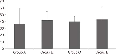
Fig. 3 Removal torque and standard deviation.
Cited by 1 articles
-
Comparison of removal torques between laser-etched and modified sandblasted acid-etched Ti implant surfaces in rabbit tibias
Kyung-Soon Park, Abdel Ghani Ibrahim Al Awamleh, Sung-Am Cho
J Adv Prosthodont. 2018;10(1):73-78. doi: 10.4047/jap.2018.10.1.73.
Reference
-
1. Marx RE, Carlson ER, Eichstaedt RM, Schimmele SR, Strauss JE, Georgeff KR. Platelet-rich plasma: Growth factor enhancement for bone grafts. Oral Surg Oral Med Oral Pathol Oral Radiol Endod. 1998; 85:638–646.2. Dohan DM, Choukroun J, Diss A, Dohan SL, Dohan AJ, Mouhyi J, Gogly B. Platelet-rich fibrin (PRF): a second-generation platelet concentrate. Part I: technological concepts and evolution. Oral Surg Oral Med Oral Pathol Oral Radiol Endod. 2006; 101:e37–e44.3. Cho SA, Lee BK, Park SH, Ahn JJ. The bone integration effects of platelet-rich fibrin by removal torque of titanium screw in rabbit tibia. Platelets. 2014; 25:562–566.4. Su CY, Kuo YP, Tseng YH, Su CH, Burnouf T. In vitro release of growth factors from platelet-rich fibrin (PRF): a proposal to optimize the clinical applications of PRF. Oral Surg Oral Med Oral Pathol Oral Radiol Endod. 2009; 108:56–61.5. Rodella LF, Favero G, Boninsegna R, Buffoli B, Labanca M, Scarì G, Sacco L, Batani T, Rezzani R. Growth factors, CD34 positive cells, and fibrin network analysis in concentrated growth factors fraction. Microsc Res Tech. 2011; 74:772–777.6. Aghaloo TL, Moy PK, Freymiller EG. Evaluation of platelet-rich plasma in combination with anorganic bovine bone in the rabbit cranium: a pilot study. Int J Oral Maxillofac Implants. 2004; 19:59–65.7. Anitua E. Plasma rich in growth factors: preliminary results of use in the preparation of future sites for implants. Int J Oral Maxillofac Implants. 1999; 14:529–535.8. Froum SJ, Wallace SS, Tarnow DP, Cho SC. Effect of platelet-rich plasma on bone growth and osseointegration in human maxillary sinus grafts: three bilateral case reports. Int J Periodontics Restorative Dent. 2002; 22:45–53.9. Mazor Z, Peleg M, Garg AK, Luboshitz J. Platelet-rich plasma for bone graft enhancement in sinus floor augmentation with simultaneous implant placement: patient series study. Implant Dent. 2004; 13:65–72.10. Whitman DH, Berry RL, Green DM. Platelet gel: an autologous alternative to fibrin glue with applications in oral and maxillofacial surgery. J Oral Maxillofac Surg. 1997; 55:1294–1299.11. Wiltfang J, Kloss FR, Kessler P, Nkenke E, Schultze-Mosgau S, Zimmermann R, Schlegel KA. Effects of platelet-rich plasma on bone healing in combination with autogenous bone and bone substitutes in critical-size defects. An animal experiment. Clin Oral Implants Res. 2004; 15:187–193.12. Picraux ST, Pope LE. Tailored surface modification by ion implantation and laser treatment. Science. 1984; 226:615–622.13. Gaggl A, Schultes G, Müller WD, Kärcher H. Scanning electron microscopical analysis of laser-treated titanium implant surfaces--a comparative study. Biomaterials. 2000; 21:1067–1073.14. Cho SA, Jung SK. A removal torque of the laser-treated titanium implants in rabbit tibia. Biomaterials. 2003; 24:4859–4863.15. Buser D, Schenk RK, Steinemann S, Fiorellini JP, Fox CH, Stich H. Influence of surface characteristics on bone integration of titanium implants. A histomorphometric study in miniature pigs. J Biomed Mater Res. 1991; 25:889–902.16. Ahn SJ, Leesungbok R, Lee SW. Histomorphometric analysis and removal torque of small diameter implants with alternative surface treatments and different designs. J Oral Implantol. 2010; 36:263–272.17. Buser D, Broggini N, Wieland M, Schenk RK, Denzer AJ, Cochran DL, Hoffmann B, Lussi A, Steinemann SG. Enhanced bone apposition to a chemically modified SLA titanium surface. J Dent Res. 2004; 83:529–533.18. Zhao G, Schwartz Z, Wieland M, Rupp F, Geis-Gerstorfer J, Cochran DL, Boyan BD. High surface energy enhances cell response to titanium substrate microstructure. J Biomed Mater Res A. 2005; 74:49–58.19. Rupp F, Scheideler L, Olshanska N, de Wild M, Wieland M, Geis-Gerstorfer J. Enhancing surface free energy and hydrophilicity through chemical modification of microstructured titanium implant surfaces. J Biomed Mater Res A. 2006; 76:323–334.20. Alissa R, Esposito M, Horner K, Oliver R. The influence of platelet-rich plasma on the healing of extraction sockets: an explorative randomised clinical trial. Eur J Oral Implantol. 2010; 3:121–134.21. Arenaz-Búa J, Luaces-Rey R, Sironvalle-Soliva S, Otero-Rico A, Charro-Huerga E, Patiño-Seijas B, García-Rozado A, Ferreras-Granados J, Vázquez-Mahía I, Lorenzo-Franco F, Martín-Sastre R, López-Cedrún JL. A comparative study of platelet-rich plasma, hydroxyapatite, demineralized bone matrix and autologous bone to promote bone regeneration after mandibular impacted third molar extraction. Med Oral Patol Oral Cir Bucal. 2010; 15:e483–e489.22. Gürbüzer B, Pikdöken L, Urhan M, Süer BT, Narin Y. Scintigraphic evaluation of early osteoblastic activity in extraction sockets treated with platelet-rich plasma. J Oral Maxillofac Surg. 2008; 66:2454–2460.23. Albanese A, Licata ME, Polizzi B, Campisi G. Platelet-rich plasma (PRP) in dental and oral surgery: from the wound healing to bone regeneration. Immun Ageing. 2013; 10:23.24. Garcia RV, Gabrielli MA, Hochuli-Vieira E, Spolidorio LC, Filho JG, Neto FA, de Cardoso LA, Shibli JA. Effect of platelet-rich plasma on peri-implant bone repair: a histologic study in dogs. J Oral Implantol. 2010; 36:281–290.25. Daif ET. Effect of autologous platelet-rich plasma on bone regeneration in mandibular fractures. Dent Traumatol. 2013; 29:399–403.26. Poeschl PW, Ziya-Ghazvini F, Schicho K, Buchta C, Moser D, Seemann R, Ewers R, Schopper C. Application of platelet-rich plasma for enhanced bone regeneration in grafted sinus. J Oral Maxillofac Surg. 2012; 70:657–664.27. Jeong IY, Cho SA. The bone healing effects of concentrated growth factors with a tooth ash by removal torque of titanium screw in rabbit tibia. Archives of Oral Biology (In review).28. Esposito M, Grusovin MG, Rees J, Karasoulos D, Felice P, Alissa R, Worthington HV, Coulthard P. Interventions for replacing missing teeth: augmentation procedures of the maxillary sinus. Cochrane Database Syst Rev. 2010; (3):CD008397.29. Mustafa K, Wennerberg A, Wroblewski J, Hultenby K, Lopez BS, Arvidson K. Determining optimal surface roughness of TiO(2) blasted titanium implant material for attachment, proliferation and differentiation of cells derived from human mandibular alveolar bone. Clin Oral Implants Res. 2001; 12:515–525.30. Rupp F, Scheideler L, Eichler M, Geis-Gerstorfer J. Wetting behavior of dental implants. Int J Oral Maxillofac Implants. 2011; 26:1256–1266.
- Full Text Links
-
- Actions
-
Cited
- CITED
-
- Close
- Share
-
- Similar articles
-
- Stability and Efficacy of Titanium in Osteointegration in a Rabbit Model
- The effects of saline soaking on the removal torque of titanium implants in rabbit tibia after 10 days
- Bone-to-Implant Contact according to the Surface Roughness of the Implants
- The effect of various surface treatment methods on the osseointegration
- CONFOCAL LASER SCANNING MICROSCOPY STUDY ON INTERFACE BONE AND TITANIUM IMPLANT COATED BY CHITOSAN
