Osteoporosis prediction from the mandible using cone-beam computed tomography
- Affiliations
-
- 1Department of Oral Medicine, Faculty of Dentistry, Damascus University, Damascus, Syria. imadbarn@gmail.com
- 2Department of Periodontology, Faculty of Dentistry, Damascus University, Damascus, Syria.
- KMID: 1974494
- DOI: http://doi.org/10.5624/isd.2014.44.4.263
Abstract
- PURPOSE
This study aimed to evaluate the use of dental cone-beam computed tomography (CBCT) in the diagnosis of osteoporosis among menopausal and postmenopausal women by using only a CBCT viewer program.
MATERIALS AND METHODS
Thirty-eight menopausal and postmenopausal women who underwent dual-energy X-ray absorptiometry (DXA) examination for hip and lumbar vertebrae were scanned using CBCT (field of view: 13 cmx15 cm; voxel size: 0.25 mm). Slices from the body of the mandible as well as the ramus were selected and some CBCT-derived variables, such as radiographic density (RD) as gray values, were calculated as gray values. Pearson's correlation, one-way analysis of variance (ANOVA), and accuracy (sensitivity and specificity) evaluation based on linear and logistic regression were performed to choose the variable that best correlated with the lumbar and femoral neck T-scores.
RESULTS
RD of the whole bone area of the mandible was the variable that best correlated with and predicted both the femoral neck and the lumbar vertebrae T-scores; further, Pearson's correlation coefficients were 0.5/0.6 (p value=0.037/0.009). The sensitivity, specificity, and accuracy based on the logistic regression were 50%, 88.9%, and 78.4%, respectively, for the femoral neck, and 46.2%, 91.3%, and 75%, respectively, for the lumbar vertebrae.
CONCLUSION
Lumbar vertebrae and femoral neck osteoporosis can be predicted with high accuracy from the RD value of the body of the mandible by using a CBCT viewer program.
MeSH Terms
Figure
-
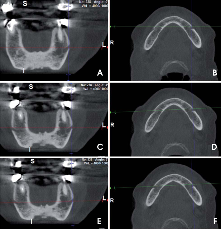
Fig. 1 The angulation adjustment procedure of the CBCT images is seen. Coronal (A) and axial (B) slices before angulation adjustment. Note that the openings of the left and the right foramina do not appear symmetrical in the same coronal slice. The angulation of the coronal slice (C) is adjusted using the coronal (the green) cursor on the axial slice window (D). Now, both foramina appear simultaneously on the coronal views. This is checked further by navigating through all the coronal slices. Using the axial (the red) cursor on the coronal slice window (E), we adjusted the angulation of the axial slice (F) to be parallel with the lower border of both foramina.
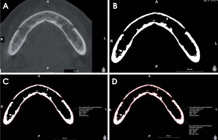
Fig. 2 Measurement procedure for the radiographic density (gray values) and the area (mm2) of the whole bony region and for the trabecular bone alone the mandibular body slice. A. The image shows the slice before the adjustment of the window width and level. B. The window width and level are adjusted to 0 and 960 gray values, respectively. C. The radiographic density (gray values) and the area (mm2) of the whole bony region are measured using the software. D. The radiographic density (gray values) and the area(mm2) of the trabecular bony region are measured.
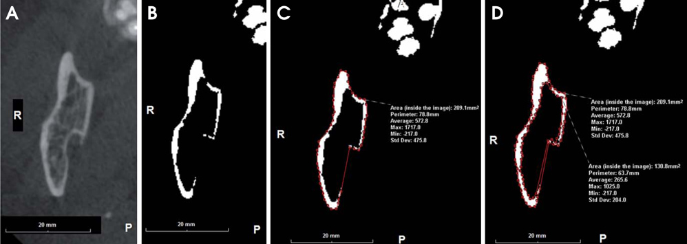
Fig. 3 Measurement procedure for the radiographic density (gray values) and the area(mm2) of the whole bony region and for the trabecular bone alone the ramus slice A. The axial image shows the slice before the adjustment of the window width and level. B. The window width and level are adjusted to 0 and 960 gray values, respectively. C. The radiographic density (gray values) and the area (mm2) of the whole bony region are measured. D. The radiographic density (gray values) and the area (mm2) of the trabecular bony region are measured.
Cited by 1 articles
-
IDIOS: An innovative index for evaluating dental imaging-based osteoporosis screening indices
Imad Barngkgei, Esam Halboub, Abeer Abdulkareem Almashraqi, Razan Khattab, Iyad Al Haffar
Imaging Sci Dent. 2016;46(3):185-202. doi: 10.5624/isd.2016.46.3.185.
Reference
-
1. NIH Consensus Development Panel on Osteoporosis Prevention Diagnosis and Therapy. Osteoporosis prevention, diagnosis, and therapy. JAMA. 2001; 285:785–795.2. Riera-Espinoza G. Epidemiology of osteoporosis in Latin America 2008. Salud Publica Mex. 2009; 51:Suppl 1. S52–S55.
Article3. Johnell O, Kanis J. Epidemiology of osteoporotic fractures. Osteoporos Int. 2005; 16:Suppl 2. S3–S7.
Article4. Lindsay R, Pack S, Li Z. Longitudinal progression of fracture prevalence through a population of postmenopausal women with osteoporosis. Osteoporos Int. 2005; 16:306–312.
Article5. Guglielmi G. Preface. Imaging of osteoporosis. Radiol Clin North Am. 2010; 48:xv.6. Nackaerts O, Jacobs R, Devlin H, Pavitt S, Bleyen E, Yan B, et al. Osteoporosis detection using intraoral densitometry. Dentomaxillofac Radiol. 2008; 37:282–287.
Article7. Verheij JG, Geraets WG, van der Stelt PF, Horner K, Lindh C, Nicopoulou-Karayianni K, et al. Prediction of osteoporosis with dental radiographs and age. Dentomaxillofac Radiol. 2009; 38:431–437.
Article8. Roberts M, Yuan J, Graham J, Jacobs R, Devlin H. Changes in mandibular cortical width measurements with age in men and women. Osteoporos Int. 2011; 22:1915–1925.
Article9. White SC, Pharoah MJ. The evolution and application of dental maxillofacial imaging modalities. Dent Clin North Am. 2008; 52:689–705.
Article10. Mozzo P, Procacci C, Tacconi A, Martini PT, Andreis IA. A new volumetric CT machine for dental imaging based on the cone-beam technique: preliminary results. Eur Radiol. 1998; 8:1558–1564.
Article11. Hua Y, Nackaerts O, Duyck J, Maes F, Jacobs R. Bone quality assessment based on cone beam computed tomography imaging. Clin Oral Implants Res. 2009; 20:767–771.
Article12. Assessment of fracture risk and its application to screening for postmenopausal osteoporosis. Report of a WHO Study Group. World Health Organ Tech Rep Ser. 1994; 843:1–129.13. Sennerby L, Andersson P, Pagliani L, Giani C, Moretti G, Molinari M, et al. Evaluation of a novel cone beam computed tomography scanner for bone density examinations in preoperative 3D reconstructions and correlation with primary implant stability. Clin Implant Dent Relat Res. (in press).
Article14. Naitoh M, Kurosu Y, Inagaki K, Katsumata A, Noguchi T, Ariji E. Assessment of mandibular buccal and lingual cortical bones in postmenopausal women. Oral Surg Oral Med Oral Pathol Oral Radiol Endod. 2007; 104:545–550.
Article15. Landis JR, Koch GG. The measurement of observer agreement for categorical data. Biometrics. 1977; 33:159–174.
Article16. Stone KL, Seeley DG, Lui LY, Cauley JA, Ensrud K, Browner WS, et al. BMD at multiple sites and risk of fracture of multiple types: long-term results from the Study of Osteoporotic Fractures. J Bone Miner Res. 2003; 18:1947–1954.
Article17. Kasturi GC, Cifu DX, Adler RA. A review of osteoporosis: part I. Impact, pathophysiology, diagnosis and unique role of the physiatrist. PM R. 2009; 1:254–260.
Article18. Genant HK, Engelke K, Prevrhal S. Advanced CT bone imaging in osteoporosis. Rheumatology (Oxford). 2008; 47:Suppl 4. iv9–iv16.
Article19. Cummings SR, Melton LJ. Epidemiology and outcomes of osteoporotic fractures. Lancet. 2002; 359:1761–1767.
Article20. Kado DM, Browner WS, Palermo L, Nevitt MC, Genant HK, Cummings SR. Vertebral fractures and mortality in older women: a prospective study. Study of Osteoporotic Fractures Research Group. Arch Intern Med. 1999; 159:1215–1220.21. Naitoh M, Katsumata A, Mitsuya S, Kamemoto H, Ariji E. Measurement of mandibles with microfocus x-ray computerized tomography and compact computerized tomography for dental use. Int J Oral Maxillofac Implants. 2004; 19:239–246.22. Cassetta M, Stefanelli LV, Di Carlo S, Pompa G, Barbato E. The accuracy of CBCT in measuring jaws bone density. Eur Rev Med Pharmacol Sci. 2012; 16:1425–1429.23. Mah P, Reeves TE, McDavid WD. Deriving Hounsfield units using grey levels in cone beam computed tomography. Dentomaxillofac Radiol. 2010; 39:323–335.
Article24. Parsa A, Ibrahim N, Hassan B, Motroni A, van der Stelt P, Wismeijer D. Reliability of voxel gray values in cone beam computed tomography for preoperative implant planning assessment. Int J Oral Maxillofac Implants. 2012; 27:1438–1442.25. Arisan V, Karabuda ZC, Avsever H, Ozdemir T. Conventional multi-slice computed tomography (CT) and cone-beam CT (CBCT) for computer-assisted implant placement. Part I: relationship of radiographic gray density and implant stability. Clin Implant Dent Relat Res. 2013; 15:893–906.
Article26. Pauwels R, Nackaerts O, Bellaiche N, Stamatakis H, Tsiklakis K, Walker A, et al. Variability of dental cone beam CT grey values for density estimations. Br J Radiol. 2013; 86:20120135.
Article27. Reeves TE, Mah P, McDavid WD. Deriving Hounsfield units using grey levels in cone beam CT: a clinical application. Dentomaxillofac Radiol. 2012; 41:500–508.
Article28. Parsa A, Ibrahim N, Hassan B, van der Stelt P, Wismeijer D. Bone quality evaluation at dental implant site using multislice CT, micro-CT, and cone beam CT. Clin Oral Implants Res. (in press).
Article29. Nomura Y, Watanabe H, Honda E, Kurabayashi T. Reliability of voxel values from cone-beam computed tomography for dental use in evaluating bone mineral density. Clin Oral Implants Res. 2010; 21:558–562.
Article30. Buyukkaplan US, Guldag MU, Yildiz M, Gumus BA. Comparison of mandibular bone mineral density in osteoporotic, osteopenic and normal elderly edentulous subjects measured by the dual-energy X-ray absorptiometry technique. Gerodontology. 2012; 29:e1098–e1102.
Article31. Carballido-Gamio J, Majumdar S. Clinical utility of microarchitecture measurements of trabecular bone. Curr Osteoporos Rep. 2006; 4:64–70.
Article32. Genant HK, Delmas PD, Chen P, Jiang Y, Eriksen EF, Dalsky GP, et al. Severity of vertebral fracture reflects deterioration of bone microarchitecture. Osteoporos Int. 2007; 18:69–76.
Article33. Veigel E, Moore RJ, Zarrinkalam MR, Schulze D, Sauerbier S, Schmelzeisen R, et al. Osteopenia in the maxillofacial area: a study in sheep. Osteoporos Int. 2011; 22:1115–1121.
Article34. van der Linden JC, Weinans H. Effects of microarchitecture on bone strength. Curr Osteoporos Rep. 2007; 5:56–61.
Article35. Singh M, Nagrath AR, Maini PS. Changes in trabecular pattern of the upper end of the femur as an index of osteoporosis. J Bone Joint Surg Am. 1970; 52:457–467.
Article36. Koh KJ, Kim KA. Utility of the computed tomography indices on cone beam computed tomography images in the diagnosis of osteoporosis in women. Imaging Sci Dent. 2011; 41:101–106.
Article37. Klemetti E, Kolmakov S, Heiskanen P, Vainio P, Lassila V. Panoramic mandibular index and bone mineral densities in postmenopausal women. Oral Surg Oral Med Oral Pathol. 1993; 75:774–779.
Article38. Jagelaviciene E, Kubilius R, Krasauskiene A. The relationship between panoramic radiomorphometric indices of the mandible and calcaneus bone mineral density. Medicina (Kaunas). 2010; 46:95–103.39. Taguchi A, Tsuda M, Ohtsuka M, Kodama I, Sanada M, Nakamoto T, et al. Use of dental panoramic radiographs in identifying younger postmenopausal women with osteoporosis. Osteoporos Int. 2006; 17:387–394.
Article40. Marquezan M, Lau TC, Mattos CT, Cunha AC, Nojima LI, Sant'Anna EF, et al. Bone mineral density. Angle Orthod. 2012; 82:62–66.
Article41. Issever AS, Link TM, Kentenich M, Rogalla P, Burghardt AJ, Kazakia GJ, et al. Assessment of trabecular bone structure using MDCT: comparison of 64- and 320-slice CT using HRpQCT as the reference standard. Eur Radiol. 2010; 20:458–468.
Article42. Norton MR, Gamble C. Bone classification: an objective scale of bone density using the computerized tomography scan. Clin Oral Implants Res. 2001; 12:79–84.
Article43. Nackaerts O, Jacobs R, Horner K, Zhao F, Lindh C, Karayianni K, et al. Bone density measurements in intra-oral radiographs. Clin Oral Investig. 2007; 11:225–229.
Article44. Damilakis J, Maris TG, Karantanas AH. An update on the assessment of osteoporosis using radiologic techniques. Eur Radiol. 2007; 17:1591–1602.
Article45. Adams JE. Advances in bone imaging for osteoporosis. Nat Rev Endocrinol. 2013; 9:28–42.
Article46. Eckstein F, Lochmuller EM, Lill CA, Kuhn V, Schneider E, Delling G, et al. Bone strength at clinically relevant sites displays substantial heterogeneity and is best predicted from site-specific bone densitometry. J Bone Miner Res. 2002; 17:162–171.
Article
- Full Text Links
-
- Actions
-
Cited
- CITED
-
- Close
- Share
-
- Similar articles
-
- Cone beam CT findings of retromolar canals: Report of cases and literature review
- Stafne bone cavity and cone-beam computed tomography: a report of two cases
- Accessory mental foramen: A rare anatomical variation detected by cone-beam computed tomography
- Management of root canal perforation by using cone-beam computed tomography
- Evaluation of imaging reformation with cone beam computed tomography for the assessment of bone density and shape in mandible
