A Case of Retroperitoneal Paraganglioma Manifested as Intractable Constipation with Paralytic Ileus and Aggravated Hyperglycemia
- Affiliations
-
- 1Department of Endocrinology and Metabolism, Kyung Hee University School of Medicine, Seoul, Korea.
- 2Research Institute of Endocrinology, Kyung Hee University School of Medicine, Seoul, Korea.
- KMID: 1966000
- DOI: http://doi.org/10.3803/jkes.2008.23.6.450
Abstract
- Paragangliomas are extra-adrenal pheochromocytomas that arise from specialized neural crest cells. They are distributed anywhere from the upper neck to the pelvic floor, and they are classified on the basis of their anatomic origin. Functioning paragangliomas can cause the same clinical manifestations as pheochromocytoma, such as hypertension, diabetes mellitus, hyperadrenergic spells and so on. We experienced a retroperitoneal paraganglioma that was found in 66 year-old male who suffered from intractable constipation, and his constipation was caused by paralytic ileus and uncontrolled hyperglycemia. After he was diagnosed, removal of the paraganglioma was done and his clinical symptoms and sustained hyperglycemia were successfully resolved.
Keyword
MeSH Terms
Figure
-

Fig. 1 Simple abdominal X-ray finding of paralytic ileus. Simple abdomen X-ray photographs shows markedly gas-distended colon with air-fluid level of stepladder pattern. It means severe paralytic ileus.

Fig. 2 CT scan finding of paraganglioma. A. Horizontal section. B. Coronal section. Two abdominal CT scan photographs show inhomogenously enhanced hypervascular mass (4.7 × 4.2 × 5.2 cm) with clear boundaries and lobulating contour located in left paraaortic area, L3 level. It contains septa-like structures and less enhanced portions with peripheral rim enhancement.

Fig. 3 I123 MIBG scan finding of paraganglioma. I123 MIBG scan photograph shows a lesion of increased uptake of radio-active materials located in left para-aortic area, suggestive of extraadrenal pheochromocytoma.

Fig. 4 Gross and microscopic findings of paraganglioma. A. It is a well defined solid mass, measuring 7.8×5×4.5 cm in dimension. Cut surface shows mahogany-colored soft and friable tumor mass associated with hemorrhage and necrosis. The mass is surrounded by thin membranous capsule. B. Some polygonal tumor cell nests make clusters resembling "Zellballen" appearance (H-E stain, ×40). C. Tumor cell has abundant granular eosinophilic cytoplasm with indistinct cell borders (H-E stain, ×100).
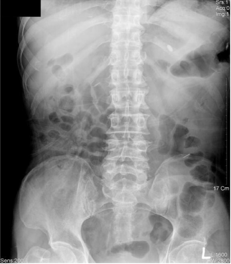
Fig. 5 Simple abdomen x-ray (post-operation follow up). Post-operation follow up simple abdomen photographs shows decreased colonic distension and bowel gas compared with previous x-ray photographs. It means improvement of the paralytic ileus.
Reference
-
1. Landsberg L, Young JB. Kasper D, Braunwald E, Fauci A, Hauser S, Longo D, Jameson L, editors. Pheochromocytoma. Harrison's principles of internal medicine. 2004. 16th Ed. New York: McGraw-Hill;2148–2152.2. Salazar A, Naik A, Rolston D. Intestinal pseudoobstruction as a presenting feature of a pheochromocytoma. J Clin Gastroenterol. 2001. 33:253–254.3. Lenz T, Gossmann J, Schulte K, Salewski L, Geiger H. Diagnosis of pheochromocytoma. Clin Lab. 2002. 48:5–18.4. Browers F, Lenders J, Eisenhofer G, Pacak K. Pheochromocytoma as an endocrine emergency. Rev Endocr Metab Disord. 2003. 4:121–128.5. Lonser RR, Glenn GM, Walther M, Chew EY, Libutti SK, Linehan WM, Oldfield EH. von Hippel-Lindau disease. Lancet. 2003. 361:2059–2067.6. Neumann HP, Berger DP, Sigmund G, Blum U, Schmidt D, Parmer RJ, Volk B, Kirste G. Pheochromocytomas, multiple endocrine neoplasia type 2, and von Hippel-Lindau disease. N Engl J Med. 1993. 329:1531–1538.7. Walther MM, Herring J, Enquist E, Keiser HR, Linehan WM. von Recklinghausen's disease and pheochromocytomas. J Urol. 1999. 162:1582–1586.8. Neumann HP, Bausch B, McWhinney SR, Bender BU, Gimm O, Franke G, Schipper J, Klisch J, Altehoefer C, Zerres K, Januszewicz A, Eng C, Smith WM, Munk R, Manz T, Glaesker S, Apel TW, Treier M, Reineke M, Walz MK, Hoang-Vu C, Brauckhoff M, Klein-Franke A, Klose P, Schmidt H, Maier-Woelfle M, Peçzkowska M, Szmigielski C, Eng C. Germ-line mutations in nonsyndromic pheochromocytoma. N Engl J Med. 2002. 346:1459–1466.9. Hayes W, Davidson A, Grimley P, Hartman D. Extraadrenal retroperitoneal paraganglioma: clinical, pathologic, and CT findings. Am J Roentgenol. 1990. 155:1247–1250.10. Kim WY, Kang KJ, Lim TJ. Functioning paraganglioma manifested cerebral hemorrhage and combined with coronary arteriovenous fistula. J Korean Surg Soc. 2002. 63:517–520.11. Young WF Jr. Paragangliomas: clinical overview. Ann N Y Acad Sci. 2006. 1073:21–29.12. Sobol SM, Dailey JC. Familial multiple cervical paragangliomas: report of a kindred and review of the literature. Otolaryngol Head Neck Surg. 1990. 102:382–390.13. Kim YT, Song YK, Lee KU, Lee M, Park KC, Yu ES, Lee JH, Jin TS. A case of paraganglioma manifested as intestinal pseudo-obstruction. Korean J Intern Med. 1990. 40:708–711.14. Hahimoto Y, Motoyoshi S, Maruyama H, Sakakida M, Yano T, Yamaguchi K, Goto K, Sugihara S, Takano S, Kambara T, Shichiri M. The treatment of pheochromocytoma associated with pseudo-obstruction and perforation of the colon, hepatic failure, and DIC. Jpn J Med. 1990. 29:341–346.15. Murakami S, Okushiba S, Ohno K, Ito K, Satou K, Sugiura H, Morikawa T, Furukawa K, Kondo S, Katoh H, Nihei K. Malignant pheochromocytoma associated with pseudo-obstruction of the colon. J Gastroenterol. 2003. 38:175–180.16. Cryer P. Physiology and pathophysiology of the human sympathoadrenal neuroendocrine system. N Engl J Med. 1980. 303:436–444.17. Vance JE, Buchanan KD, O'Hara D. Insulin and glucagon responses in subjects with pheochromocytoma: Effect of alpha adrenergic blockade. J Clin Endocrinol Metab. 1969. 29:911–916.18. Stenstrom G, Sjostrom L, Smith U. Diabetes mellitus in phaeochromocytoma: fasting blood glucose levels before and after surgery in 60 patients with phaeochromocytoma. Acta Endocrinol. 1984. 106:511–515.19. Ball AB, Tait DM, Fisher C, Sinnett HD, Harmer CL. Treatment of metastatic paraganglioma by surgery, radiotherapy and I-131 MIBG. Eur J Surg Oncology. 1991. 17:543–546.20. Sisson JC, Shapiro B, Beierwaltes WH, Glowniak JV, Nakajo M, Mangner TJ, Carey JE, Swanson DP, Copp JE, Satterlee WG, Wieland DM. Radiopharmaceutical treatment of malignant pheochromocytoma. J Nucl Med. 1984. 24:197–206.
- Full Text Links
-
- Actions
-
Cited
- CITED
-
- Close
- Share
-
- Similar articles
-
- Quetiapine Related Acute Paralytic Ileus in a Bipolar I Disorder Patient with Successful Low Dose Amisulpride Substitution: A Case Report
- Non-Functional Retroperitoneal Paraganglioma Mimicking an Ovary Mass: A Case Report
- Fine Needle Aspiration Cytology of Retroperitoneal Paraganglioma with an Unusual Pattern: A Case Report
- A Case of Retroperitoneal Paraganglioma Accompanying Bile Duct Dilatation
- Neostigmine Treatment of Paralytic Ileus in Critically Ill Patients
