Unilateral intraoral vertical ramus osteotomy based on preoperative three-dimensional simulation surgery in a patient with facial asymmetry
- Affiliations
-
- 1Department of Oral and Maxillofacial Surgery, National Health Insurance Service Ilsan Hospital, Goyang, Korea. omfs1ksh@hanmail.net
- KMID: 1960970
- DOI: http://doi.org/10.5125/jkaoms.2014.40.1.32
Abstract
- Preoperative surgical simulation in orthognathic surgery has progressed in recent years; the movement of the mandible can be anticipated through three-dimensional (3D) simulation surgery before the actual procedure. In this case report, the mandible was moved to the intended postoperative occlusion through preoperative surgical 3D simulation. Right-side condylar movement change was very slight in the surgical simulation, suggesting the possibility of mandibular surgery that included only left-side ramal osteotomy. This case report describes a patient with a mild asymmetric facial profile in which the mandibular menton had been deviated to the right and the lips canted down to the left. Before surgery, three-dimensional surgical simulation was used to evaluate and confirm a position for the condyle as well as the symmetrical postoperative state of the face. Facial asymmetry was resolved with minimal surgical treatment through unilateral intraoral vertical ramus osteotomy on the left side of the mandible. It would be a valuable complement for the reduction of the surgical treatment if one could decide with good predictability when an isolated intraoral vertical ramus osteotomy can be done without a compensatory osteotomy on the contralateral side.
Keyword
MeSH Terms
Figure
-
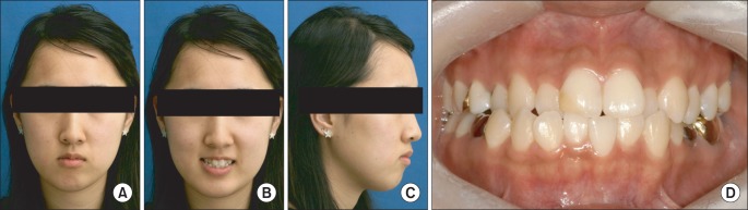
Fig. 1 A-C. Initial pretreatment extraoral photographs. D. Intraoral photograph.
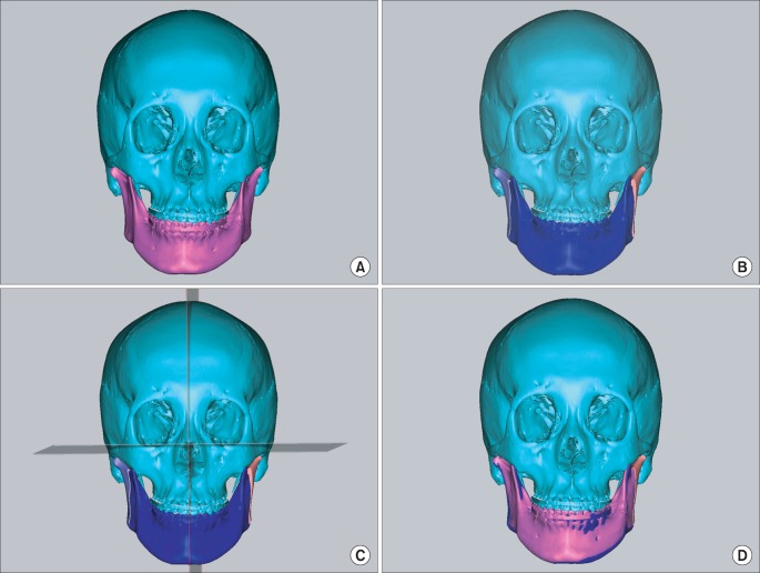
Fig. 2 Mandibular simulation surgery. A. Preoperative three-dimensional (3D) skeletal image overlapped with current occlusion digital cast images. B, C. 3D images of the mandibular setback (blue) repositioning under bilateral ramus osteotomy was simulated and evaluated according to the planned postoperative occlusion images. D. Images were overlaid consisting of the current mandible image (pink) and the repositioned mandible image (blue) in the 3D surgical simulation with the planned postoperative occlusion state.
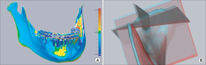
Fig. 3 A. Discrepancies were color-coded and evaluated in the three-dimensional (3D) image based on the range of difference values in the superimposed preoperative mandibular image and the simulated repositioned mandible. B. Discrepancies of the right condyle were evaluated in the 3D image based on the moved range of 3D coordinate planes in the superimposed preoperative mandibular image (sky blue) and the simulated repositioned mandible (red).
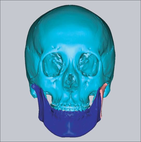
Fig. 4 Final surgical simulation image, with only left mandibular ramus osteotomy. Final surgical simulation with only left mandibular ramus osteotomy (blue) was confirmed, and decided as final surgical plan.
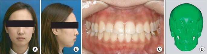
Fig. 5 Extraoral photographs (A, B), intraoral photograph (C), and three-dimensional facial computed tomography (D) of the final result.
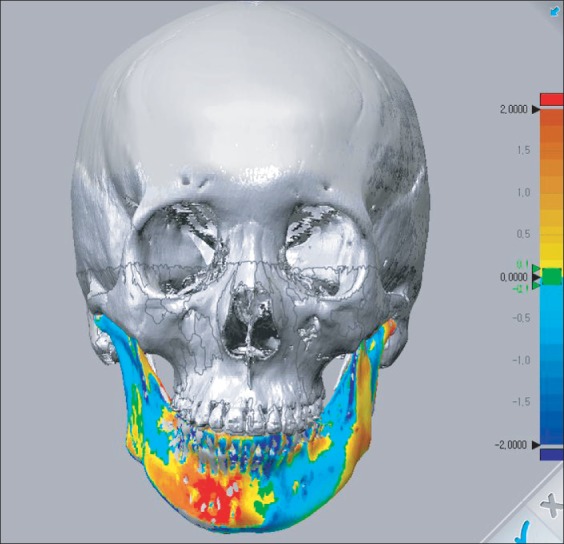
Fig. 6 Discrepancies were color-coded in the three-dimensional image based on the range of difference values in the superimposed preoperative mandibular image and the final result mandible.
Cited by 1 articles
-
Single-tooth dento-osseous osteotomy with a computer-aided design/computer-aided manufacturing surgical guide
Sang-Hoon Kang, Moon-Key Kim, Ji-Yeon Lee
J Korean Assoc Oral Maxillofac Surg. 2016;42(2):127-130. doi: 10.5125/jkaoms.2016.42.2.127.
Reference
-
1. Wohlwender I, Daake G, Weingart D, Brandstätter A, Kessler P, Lethaus B. Condylar resorption and functional outcome after unilateral sagittal split osteotomy. Oral Surg Oral Med Oral Pathol Oral Radiol Endod. 2011; 112:315–321. PMID: 21292514.
Article2. Kang SH, Kim MK, Park WS, Lee SH. Accurate computerised mandibular simulation in orthognathic surgery: a new method for integrating the planned postoperative occlusion model. Br J Oral Maxillofac Surg. 2010; 48:305–307. PMID: 19616350.
Article3. Kang SH, Lee JW, Kim MK. Use of the surface-based registration function of computer-aided design/computer-aided manufacturing software in medical simulation software for three-dimensional simulation of orthognathic surgery. J Korean Assoc Oral Maxillofac Surg. 2013; 39:197–199. PMID: 24471043.
Article4. Rubens BC, Stoelinga PJ, Weaver TJ, Blijdorp PA. Management of malunited mandibular condylar fractures. Int J Oral Maxillofac Surg. 1990; 19:22–25. PMID: 2110955.
Article5. Ueki K, Marukawa K, Shimada M, Hashiba Y, Nakgawa K, Yamamoto E. Condylar and disc positions after sagittal split ramus osteotomy with and without Le Fort I osteotomy. Oral Surg Oral Med Oral Pathol Oral Radiol Endod. 2007; 103:342–348. PMID: 17321444.
Article6. Al-Gunaid T, Yamada K, Takagi R, Saito C, Saito I. Postoperative stability of bimaxillary surgery in Class III patients with mandibular protrusion and mandibular deviation: a frontal cephalometric study. Int J Oral Maxillofac Surg. 2008; 37:992–998. PMID: 18621507.
Article7. Ueki K, Moroi A, Sotobori M, Ishihara Y, Marukawa K, Yoshizawa K, et al. Changes in temporomandibular joint and ramus after sagittal split ramus osteotomy in mandibular prognathism patients with and without asymmetry. J Craniomaxillofac Surg. 2012; 40:821–827. PMID: 22507292.
Article8. Fang B, Shen GF, Yang C, Wu Y, Feng YM, Mao LX, et al. Changes in condylar and joint disc positions after bilateral sagittal split ramus osteotomy for correction of mandibular prognathism. Int J Oral Maxillofac Surg. 2009; 38:726–730. PMID: 19375280.
Article9. Zhao Q, Hu J, Wang D, Zhu S. Changes in the temporomandibular joint after mandibular setback surgery in monkeys: intraoral vertical versus sagittal split ramus osteotomy. Oral Surg Oral Med Oral Pathol Oral Radiol Endod. 2007; 104:329–337. PMID: 17428700.
Article10. Hashimoto T, Fukunaga T, Kuroda S, Sakai Y, Yamashiro T, Takano-Yamamoto T. Mandibular deviation and canted maxillary occlusal plane treated with miniscrews and intraoral vertical ramus osteotomy: functional and morphologic changes. Am J Orthod Dentofacial Orthop. 2009; 136:868–877. PMID: 19962611.
Article
- Full Text Links
-
- Actions
-
Cited
- CITED
-
- Close
- Share
-
- Similar articles
-
- Facial nerve injury related to the intraoral vertical ramus osteotomy: A case report
- Unilateral intraoral vertical ramus osteotomy and sagittal split ramus osteotomy for the treatment of asymmetric mandibles
- Unilateral bimaxillary vertical elongation by maxillary distraction osteogenesis and mandibular sagittal split ramus osteotomy: a case report
- Sagittal split ramus osteotomy, intraoral vertical ramus osteotomy, and lateral corticectomy for asymmetric mandibular prognathism
- A study on the prediction of orthognathic surgery using rapid prototyping model technology
