Plexiform Angiomyxoid Myofibroblastic Tumor of the Stomach: Report of a Case and Review of the Literature
- Affiliations
-
- 1Department of Radiology, Haeundae Paik Hospital, Inje University College of Medicine, Busan, Korea. radyjh@hanmail.net
- 2Department of Pathology, Haeundae Paik Hospital, Inje University College of Medicine, Busan, Korea.
- KMID: 1897260
- DOI: http://doi.org/10.3348/jksr.2014.70.1.47
Abstract
- We report a case of a plexiform angiomyxoid myofibroblastic tumor of the stomach that developed in a 38-year-old woman who underwent gastrofiberscopy and multi-detector computed tomography scans due to dyspepsia for 3 months. There was a subepithelial protruding mass with a small central mucosal ulceration and heterogeneous prominent enhancement in the gastric upper body along the greater curvature. The tumor measured 3.5 x 2.3 cm in size and showed a multinodular plexiform growth pattern of bland spindle cells in the myxoid stroma with abundant small blood vessels. The tumor cells were negative for CD117 (c-KIT), CD34, and S-100 protein but diffusely positive for smooth muscle actin and Alcian blue. The pathologic examination demonstrated that the lesion was a plexiform angiomyxoid myofibroblastic tumor. The patient is doing well and has had no recurrence or metastasis for 6 months after the wedge resection.
MeSH Terms
Figure
-

Fig. 1 A 38-year-old woman with an incidentally detected subepithelial tumor of the stomach. A-C. MDCT scan axial image at the late arterial phase (A), delayed phase (B), sagittal-reconstructed scan (C) demonstrating a 3 × 2 cm-sized sessile polypoid mass with prominent contrast enhancement in the high body along the greater curvature. The enhancement pattern is heterogeneous, with some portions demonstrating a highly vascular enhancement and some portions showing a lesser degree of enhancement. D. Upon endoscopy, a 3 × 2 cm-sized luminal protruding polypoid subepithelial tumor lesion with central mucosal ulceration is found at the upper body, posterior wall and greater curvature side of the stomach. Note.-MDCT = multi-detector computed tomography
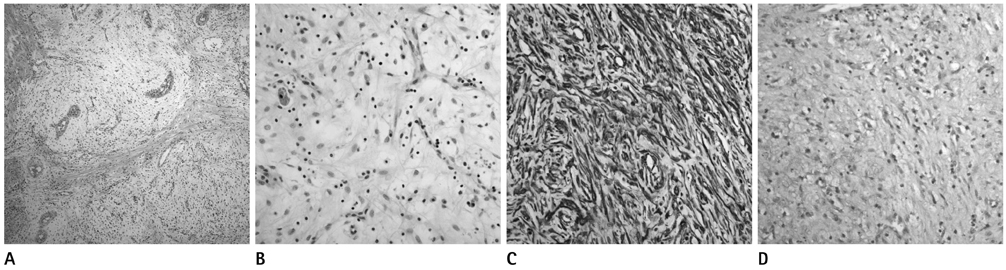
Fig. 2 A 38-year-old woman performed wedge resection for an incidentally detected subepithelial tumor of the stomach. A. Upon histopathology (Hematoxylin and Eosin, × 100), the tumor is characterized by multinodular plexiform growth pattern. B. The tumor is composed of bland-looking spindle cells scattered in the myxoid stroma rich in small vessels (Hematoxylin and Eosin, × 400). C. Immunohistochemically, the tumor cells are positive for smooth muscle actin (× 400). D. The myxoid stroma is positive under alcian blue staining (× 400).
Cited by 1 articles
-
Gastric Plexiform Fibromyxoma Incidentally Found in the Routine Checkup
Eun Jeong Gong, Young Joo Yang, Sang Hak Han, Chang Seok Bang
Korean J Gastroenterol. 2022;79(2):83-86. doi: 10.4166/kjg.2022.019.
Reference
-
1. Takahashi Y, Shimizu S, Ishida T, Aita K, Toida S, Fukusato T, et al. Plexiform angiomyxoid myofibroblastic tumor of the stomach. Am J Surg Pathol. 2007; 31:724–728.2. Kim A, Bae YK, Shin HC, Choi JH. Plexiform angiomyxoid myofibroblastic tumor of the stomach: a case report. J Korean Med Sci. 2011; 26:1508–1511.3. Miettinen M, Makhlouf HR, Sobin LH, Lasota J. Plexiform fibromyxoma: a distinctive benign gastric antral neoplasm not to be confused with a myxoid GIST. Am J Surg Pathol. 2009; 33:1624–1632.4. Yoshida A, Klimstra DS, Antonescu CR. Plexiform angiomyxoid tumor of the stomach. Am J Surg Pathol. 2008; 32:1910–1912. author reply 1912-1913.5. Takahashi Y, Suzuki M, Fukusato T. Plexiform angiomyxoid myofibroblastic tumor of the stomach. World J Gastroenterol. 2010; 16:2835–2840.6. Wang WY, Li JN, Li GD. Plexiform angiomyxoid myofibroblastic tumour of the gastric fundus: successful diagnosis and treatment by endoscopy. J Clin Pathol. 2010; 63:569–570.7. Kang Y, Jung W, Do IG, Lee EJ, Lee MH, Kim KM, et al. Plexiform angiomyxoid myofibroblastic tumor of the stomach: report of two cases and review of the literature. Korean J Pathol. 2012; 46:292–296.8. Sing Y, Subrayan S, Mqadi B, Ramdial PK, Reddy J, Moodley MS, et al. Gastric plexiform angiomyxoid myofibroblastic tumor. Pathol Int. 2010; 60:621–625.9. Rau TT, Hartmann A, Dietmaier W, Schmitz J, Hohenberger W, Hofstaedter F, et al. Plexiform angiomyxoid myofibroblastic tumour: differential diagnosis of gastrointestinal stromal tumour in the stomach. J Clin Pathol. 2008; 61:1136–1137.10. Galant C, Rousseau E, Ho Minh Duc DK, Pauwels P. Re: Plexiform angiomyxoid myofibroblastic tumor of the stomach. Am J Surg Pathol. 2008; 32:1910. author reply 1912-1913.11. Miettinen M, Sarlomo-Rikala M, Lasota J. Gastrointestinal stromal tumors: recent advances in understanding of their biology. Hum Pathol. 1999; 30:1213–1220.12. Levy AD, Remotti HE, Thompson WM, Sobin LH, Miettinen M. Gastrointestinal stromal tumors: radiologic features with pathologic correlation. Radiographics. 2003; 23:283–304. 456quiz 532.13. Lee MJ, Lim JS, Kwon JE, Kim H, Hyung WJ, Park MS, et al. Gastric true leiomyoma: computed tomographic findings and pathological correlation. J Comput Assist Tomogr. 2007; 31:204–208.14. Choi JW, Choi D, Kim KM, Sohn TS, Lee JH, Kim HJ, et al. Small submucosal tumors of the stomach: differentiation of gastric schwannoma from gastrointestinal stromal tumor with CT. Korean J Radiol. 2012; 13:425–433.15. Lee NK, Kim S, Kim GH, Jeon TY, Kim DH, Jang HJ, et al. Hypervascular subepithelial gastrointestinal masses: CT-pathologic correlation. Radiographics. 2010; 30:1915–1934.16. Kim JK, Won JH, Cho YK, Kim MW, Joo HJ, Suh JH. Glomus tumor of the stomach: CT findings. Abdom Imaging. 2001; 26:303–305.17. Kim JY, Lee JM, Kim KW, Park HS, Choi JY, Kim SH, et al. Ectopic pancreas: CT findings with emphasis on differentiation from small gastrointestinal stromal tumor and leiomyoma. Radiology. 2009; 252:92–100.
- Full Text Links
-
- Actions
-
Cited
- CITED
-
- Close
- Share
-
- Similar articles
-
- Plexiform Angiomyxoid Myofibroblastic Tumor of the Stomach: A Case Report
- Plexiform Angiomyxoid Myofibroblastic Tumor of the Stomach: Report of Two Cases and Review of the Literature
- Plexiform Angiomyxoid Myofibroblastic Tumor of the Stomach: a Rare Case
- Inflammatory Myofibroblastic Tumor of Nasal Septum after Septoplasty: A Case Report
- A Case of Myxoid Plexiform Fibrohistiocytic Tumor
