J Korean Neurosurg Soc.
2015 May;57(5):386-388. 10.3340/jkns.2015.57.5.386.
Unusual Position and Presentation of Frontobasal Meningoencephalocela
- Affiliations
-
- 1Department of Neurosurgery, Clinical Hospital Split, Split, Croatia. zeljko.busic1@st.htnet.hr
- 2Emergency Medical Services, Split, Croatia.
- 3Department of Maxillofacial Surgery, Clinical Hospital Split, Split, Croatia.
- KMID: 1881739
- DOI: http://doi.org/10.3340/jkns.2015.57.5.386
Abstract
- We wish to show our experiance with threating a rare congenital brain malformation-encephalocele. It is a protusion of brain matter with greater incidence in the Far East. Our case is even more curious because of the site of occurrence-frontobasal. Most of encephalocele occur in the occipital region. In this article we report a case of a 57-year-old woman, without deformations on the face, which had epileptic seizures and in spite of receiving antiepileptic drug. She was also frequently treated for sinusitis. She never had rhinoliquorrhea, nor was she diagnosed to have meningitis. In the last few years she had difficulty breathing on her right nostril. After she was diagnosed with encephalocele and treated surgically her recovery was complete and she is without the seizures.
Keyword
MeSH Terms
Figure
-
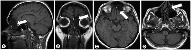
Fig. 1 MR-T1 images (A : sagital section, B : coronal section, C and D : axial section). Arrows showing meningoencephalocela.

Fig. 2 Facial appearance of our patient.
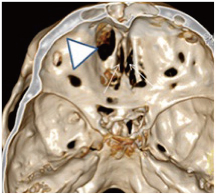
Fig. 3 CT reconstruction of the skull base; arrowhead showing skull base defect; arrows indicate the preserved lamina cribrosa.
Reference
-
1. Cho BK, Baek SH, Kim ES, Chung YS, Wang GC, Han DH. The clinical analysis of 22 cases of encephalocele. J Korean Neurosurg Soc. 1991; 20:1040–1047.2. Elster AD, Branch CL Jr. Transalar sphenoidal encephaloceles : clinical and radiologic findings. Radiology. 1989; 170(1 Pt 1):245–247. PMID: 2909104.
Article3. Mahajan C, Rath GP, Dash HH, Bithal PK. Perioperative management of children with encephalocele : an institutional experience. J Neurosurg Anesthesiol. 2011; 23:352–356. PMID: 21633311.4. Rampersaud E, Melvin EC, Speer MC. Nonsyndromic neural tube defects : genetic basis and genetic investigations. In : Wyszynsk DF, editor. Neural Tube Defects : from Origin to Treatment. New York: Oxford University Press;2006. p. 165–175.5. Shilpakar SK, Sharma MR. Surgical management of encephaloceles. J Neuroscience. 2008; 1:45–48.6. Suh MS, Hong SK, Kim HJ, Han YP. Sincipital encephalocele : report of 3 operative cases. J Korean Neurosurg Soc. 1987; 16:897–903.7. Yamashita H, Kurihara M, Kawano T, Mori K, Kunimura M. [Basal encephalomeningocele in an adult-a case report and clinico-anatomical classification]. No Shinkei Geka. 1985; 13:425–431. PMID: 4022246.
- Full Text Links
-
- Actions
-
Cited
- CITED
-
- Close
- Share
-
- Similar articles
-
- The Clinical Analysis of 22 Cases of Encephalocele
- Unusual Presentation of Secondary Syphilis in Korea: 2010-2014 Review
- Pan-vasculitis and fulminant hepatitis following routine vaccination at the age of 4 months
- Dedifferentiated Chondrosarcoma of the Rib Masquerading as a Giant Chest Wall Tumor in a Teenage Girl: An Unusual Presentation
- Hemangiopericytoma of renal sinus expanding to the renal hilum: an unusual presentation causes misinterpretation as transitional cell carcinoma
