Tuberc Respir Dis.
2012 Dec;73(6):336-341. 10.4046/trd.2012.73.6.336.
A Case of Human Herpes Virus-8 Unrelated Primary Effusion Lymphoma-Like Lymphoma Presented as Pleural Effusion
- Affiliations
-
- 1Department of Internal Medicine, CHA Bundang Medical Center, CHA University College of Medicine, Seongnam, Korea. imekkim@hanmail.net
- 2Department of Pathology, CHA Bundang Medical Center, CHA University College of Medicine, Seongnam, Korea.
- KMID: 1842928
- DOI: http://doi.org/10.4046/trd.2012.73.6.336
Abstract
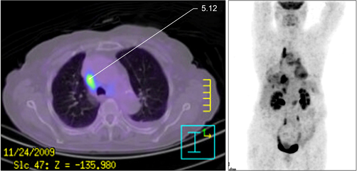
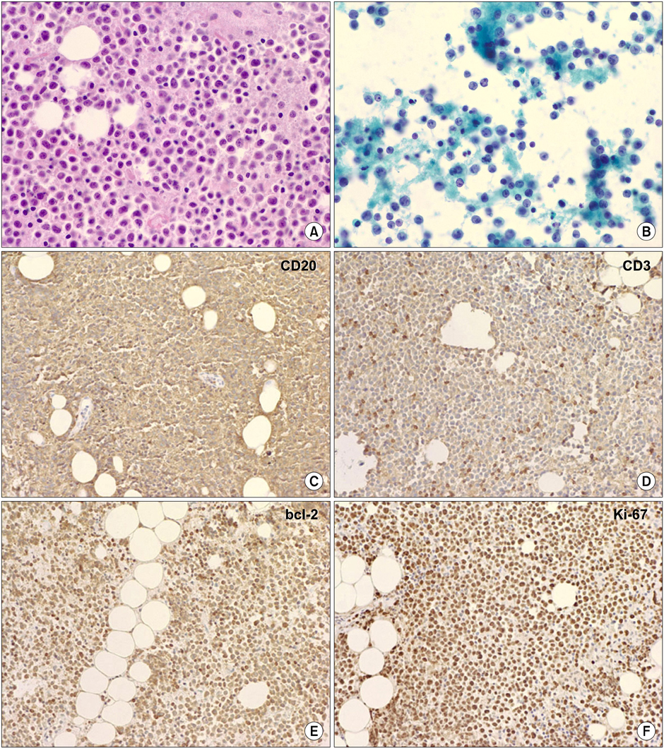
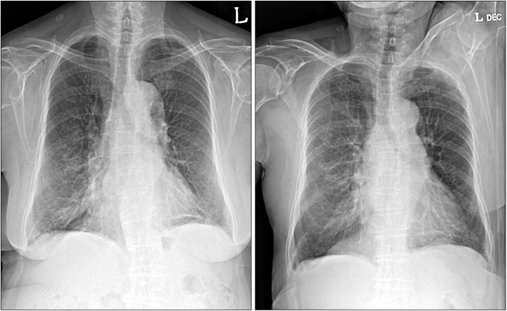
- Primary effusion lymphoma (PEL) is a rare type of lymphoma that arises in the body cavity without detectable masses. It is associated with human herpes virus-8 (HHV-8), Epstein-Barr virus (EBV), and human immunodeficiency virus (HIV). Recently, PEL unrelated to viral infection has been reported and it has been termed HHV-8 unrelated primary effusion lymphoma-like lymphoma (HHV-8 unrelated PEL-like lymphoma). Here, we report a case of HHV-8 unrelated PEL-like lymphoma in an 80-year-old woman. Chest X-ray and computed tomography revealed left-sided pleural effusion. Pleural effusion analysis and mediastinoscopic biopsy showed atypical cells that had originated from the B cells. The cells were positive for CD20 and bcl-2, but negative for CD3, CD5, CD21, CD30, CD138, epithelial membrane antigen, and HHV-8. Serological tests for HIV and EBV were negative. Considering the patient's age, further treatments were not performed. She has shown good prognosis without chemotherapy for more than 18 months.
MeSH Terms
Figure
Cited by 1 articles
-
Human Herpesvirus 8–Unrelated Primary Effusion Lymphoma–Like Lymphoma in an Elderly Korean Patient with a Good Response to Rituximab Plus Cyclophosphamide, Doxorubicin, Vincristine, and Prednisolone
Junghoon Shin, Jeong-Ok Lee, Ji-Young Choe, Soo-Mee Bang, Jong-Seok Lee
Cancer Res Treat. 2017;49(1):274-278. doi: 10.4143/crt.2016.076.
Reference
-
1. Nador RG, Cesarman E, Chadburn A, Dawson DB, Ansari MQ, Sald J, et al. Primary effusion lymphoma: a distinct clinicopathologic entity associated with the Kaposi's sarcoma-associated herpes virus. Blood. 1996. 88:645–656.2. Cesarman E, Chang Y, Moore PS, Said JW, Knowles DM. Kaposi's sarcoma-associated herpesvirus-like DNA sequences in AIDS-related body-cavity-based lymphomas. N Engl J Med. 1995. 332:1186–1191.3. Chen YB, Rahemtullah A, Hochberg E. Primary effusion lymphoma. Oncologist. 2007. 12:569–576.4. Banks PM, Warnke RA. Jaffe ES, Harris NL, Stein H, Vardiman JW, editors. Primary effusion lymphoma. World Health Organization classification of tumors: pathology and genetics of tumors of haematopoietic and lymphoid tissues. 2001. Lyon: IARC Press;179–180.5. Adiguzel C, Bozkurt SU, Kaygusuz I, Uzay A, Tecimer T, Bayik M. Human herpes virus 8-unrelated primary effusion lymphoma-like lymphoma: report of a rare case and review of the literature. APMIS. 2009. 117:222–229.6. Tanaka S, Katano H, Tsukamoto K, Jin M, Oikawa S, Nishihara H, et al. HHV8-negative primary effusion lymphoma of the peritoneal cavity presenting with a distinct immunohistochemical phenotype. Pathol Int. 2001. 51:293–300.7. Carbone A, Gloghini A. PEL and HHV8-unrelated effusion lymphomas: classification and diagnosis. Cancer. 2008. 114:225–227.8. Takao T, Kobayashi Y, Kuroda J, Omoto A, Nishimura T, Kamitsuji Y, et al. Rituximab is effective for human herpesvirus-8-negative primary effusion lymphoma with CD20 phenotype associated hepatitis C virus-related liver cirrhosis. Am J Hematol. 2004. 77:419–420.9. Matsumoto Y, Nomura K, Ueda K, Satoh K, Yasuda N, Taki T, et al. Human herpesvirus 8-negative malignant effusion lymphoma: a distinct clinical entity and successful treatment with rituximab. Leuk Lymphoma. 2005. 46:415–419.10. Ichinohasama R, Miura I, Kobayashi N, Saitoh Y, DeCoteau JF, Saiki Y, et al. Herpes virus type 8-negative primary effusion lymphoma associated with PAX-5 gene rearrangement and hepatitis C virus: a case report and review of the literature. Am J Surg Pathol. 1998. 22:1528–1537.11. Inoue S, Miyamoto T, Yoshino T, Yamadori I, Hagari Y, Yamamoto O. Primary effusion lymphoma with skin involvement. J Clin Pathol. 2006. 59:1221–1222.12. Lee YC, Rogers JT, Rodriguez RM, Miller KD, Light RW. Adenosine deaminase levels in nontuberculous lymphocytic pleural effusions. Chest. 2001. 120:356–361.13. Terasaki Y, Okumura H, Saito K, Sato Y, Yoshino T, Ichinohasama R, et al. HHV-8/KSHV-negative and CD20-positive primary effusion lymphoma successfully treated by pleural drainage followed by chemotherapy containing rituximab. Intern Med. 2008. 47:2175–2178.
- Full Text Links
- Actions
-
Cited
- CITED
-
- Close
- Share
- Similar articles
-
- Primary Effusion Lymphoma in a Non-Human Immunodeficiency Virus Patient: A Case Report
- Primary Effusion Lymphoma: An Untrivial Differential Diagnosis for Ascites
- A Case of Primary Pleural Effusion Lymphoma
- A Case of Primary Effusion Lymphoma in a Patient with Chronic Kidney Disease
- Human Herpesvirus 8–Unrelated Primary Effusion Lymphoma–Like Lymphoma in an Elderly Korean Patient with a Good Response to Rituximab Plus Cyclophosphamide, Doxorubicin, Vincristine, and Prednisolone


