Cancer Res Treat.
2008 Sep;40(3):141-144.
Hepatocellular Carcinoma Presenting as Uterine Metastasis
- Affiliations
-
- 1Department of Obstetrics and Gynecology, Chonnam National University Medical School, Gwangju, Korea. seokmo2001@hanmail.net
- 2Department of Pathology, Chonnam National University Medical School, Gwangju, Korea.
Abstract
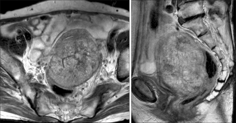
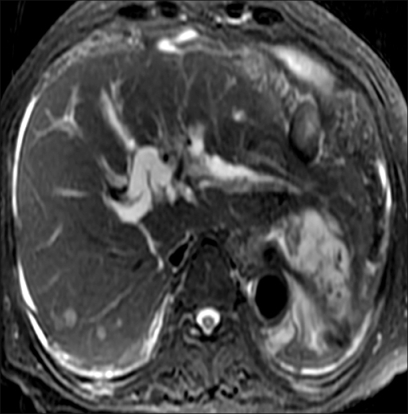
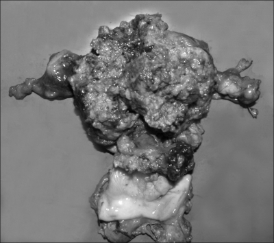
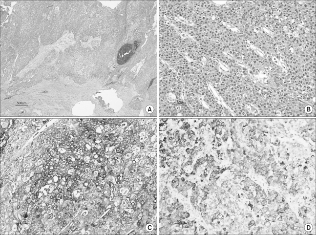
- Metastatic extragenital cancer that spreads to the uterus is rare. When it occurs, the extragenital primary disease is often in the breast or gastrointestinal tract. We report here on a case of hepatocellular carcinoma (HCC) that metastasis to the uterus. The patient was admitted for evaluation of a pelvic mass. The serum alpha-fetoprotein level was highly elevated. Magnetic resonance imaging of the abdomen and pelvis showed hepatic and uterine masses. The patient underwent surgical treatment. The histopathologic findings and immunohistochemical staining results of the uterine mass were characteristics of metastatic HCC. The endometrium and both ovaries were free of tumor. Up to now, there have been only two cases of uterine metastasis from HCC reported in the English literature. This case is the first documented instance of a metastatic uterine tumor from HCC that spared both ovaries.
MeSH Terms
Figure
Reference
-
1. Kumar NB, Hart WR. Metastases to the uterine corpus from extragenital cancers. A clinicopathologic study of 63 cases. Cancer. 1982; 50:2163–2169. PMID: 7127256.
Article2. Mazur MT, Hsueh S, Gersell DJ. Metastases to the female genital tract. Analysis of 325 cases. Cancer. 1984; 53:1978–1984. PMID: 6322966.
Article3. Stemmermann GN. Extrapelvic carcinoma metastatic to the uterus. Am J Obstet Gynecol. 1961; 82:1261–1266. PMID: 13916812.
Article4. Charache H. Metastatic carcinoma in the uterus. Am J Surg. 1941; 53:152–157.
Article5. Ryo E, Sato T, Takeshita S, Ayabe T, Tanaka F. Uterine metastasis from hepatocellular carcinoma: a case report. Int J Gynecol Cancer. 2006; 16:1894–1896. PMID: 17009988.
Article6. Okuda K, Ohtsuki T, Obata H, Tomimatsu M, Okazaki N, Hasegawa H, et al. Natural history of hepatocellular carcinoma and prognosis in relation to treatment: study of 850 patients. Cancer. 1985; 56:918–928. PMID: 2990661.
Article7. Katyal S, Oliver JH III, Peterson MS, Ferris JV, Carr BS, Baron RL. Extrahepatic metastases of hepatocellular carcinoma. Radiology. 2000; 216:698–703. PMID: 10966697.
Article8. Yeu-Tsu ML, Geer DA. Primary liver cancer: pattern of metastases. J Surg Oncol. 1987; 36:26–31. PMID: 3041113.9. Legg JW. Melanotic sarcoma of the eyeball: secondary growths in the organs of the chest and belly, particularly in the liver. Trans Pathol Soc Lond. 1878; 29:225–230.10. Takano M, Shibasaki T, Sato K, Aida S, Kikuchi Y. Malignant mixed Mullerian tumor of the uterine corpus with alpha-fetoprotein producing hepatoid adenocarcinoma component. Gynecol Oncol. 2003; 91:444–448. PMID: 14599882.11. Adams SF, Yamada SD, Montag A, Rotmensch J. An alpha fetoprotein-producing hepatoid adenocarcinoma of the endometrium. Gynecol Oncol. 2001; 83:418–421. PMID: 11606109.12. Spatz A, Bouron D, Pautier P, Castaigne D, Duvillard P. Primary yolk sac tumor of the endometrium: a case report and review of the literature. Gynecol Oncol. 1998; 70:285–288. PMID: 9740707.
Article13. Kubo K, Lee G, Yamauchi K, Kitagawa T. Alpha-fetoprotein producing papillary adenocarcinoma originating from a uterine body. Acta Pathol Jpn. 1991; 41:399–403. PMID: 1714227.
- Full Text Links
- Actions
-
Cited
- CITED
-
- Close
- Share
- Similar articles
-
- A Case of Intracardiac Metastasis of Hepatocellular Carcinoma Presenting with Functional Tricuspid Valve Stenosis Accompanied with Hepatopulmonary Syndrome
- Spinal Epidural Metastasis Presenting Atypical Images in Hepatocellular Carcinoma
- Cutaneous Metastasis from Hepatocellular Carcinoma Mimicking Pyogenic Granuloma
- Two Cases of Cutaneous Metastasis Originating from Hepatocellular Carcinoma
- Cranial Metastasis of Hepatocellular Carcinoma: Report of Three Cases


