Comparison of alveolar ridge preservation methods using three-dimensional micro-computed tomographic analysis and two-dimensional histometric evaluation
- Affiliations
-
- 1Department of Oral Anatomy, Seoul National University School of Dentistry, Seoul, Korea.
- 2Department of Periodontology and Dental Research Institute, School of Dentistry, Seoul National University, Seoul, Korea. periopf@snu.ac.kr
- 3Department of Oral and Maxillofacial Surgery, School of Dentistry, Seoul National University, Seoul, Korea.
- 4Department of Oral and Maxillofacial Radiology and Dental Research Institute, School of Dentistry, Seoul National University, Seoul, Korea.
- KMID: 1799596
- DOI: http://doi.org/10.5624/isd.2014.44.2.143
Abstract
- PURPOSE
This study evaluated the efficacy of alveolar ridge preservation methods with and without primary wound closure and the relationship between histometric and micro-computed tomographic (CT) data.
MATERIALS AND METHODS
Porcine hydroxyapatite with polytetrafluoroethylene membrane was implanted into a canine extraction socket. The density of the total mineralized tissue, remaining hydroxyapatite, and new bone was analyzed by histometry and micro-CT. The statistical association between these methods was evaluated.
RESULTS
Histometry and micro-CT showed that the group which underwent alveolar preservation without primary wound closure had significantly higher new bone density than the group with primary wound closure (P<0.05). However, there was no significant association between the data from histometry and micro-CT analysis.
CONCLUSION
These results suggest that alveolar ridge preservation without primary wound closure enhanced new bone formation more effectively than that with primary wound closure. Further investigation is needed with respect to the comparison of histometry and micro-CT analysis.
MeSH Terms
Figure
-
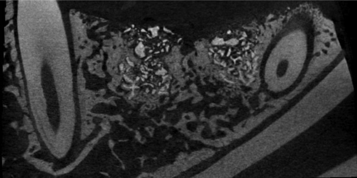
Fig. 1 Micro-computed tomograph shows radiographic image for mineral tissue measurement (19.75-µm resolution, 100 kV, 100 µA).

Fig. 2 Microphotograph shows the histologic specimen for mineral tissue measurement (H&E stain, 12.5×).
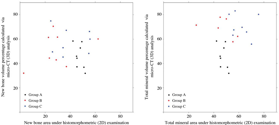
Fig. 3 A graph shows the relationship between the two-dimensional histometry and three-dimensional micro-CT data. There is no significant association between the 2D and 3D measurements in the amount of new bone formation (left, P=0.938) or total mineral content (right, P=0.200). Group A, control; group B, porcine hydroxyapatite with primary wound closure; and group C, porcine hydroxyapatite with secondary wound closure.
Cited by 1 articles
-
Three-dimensional microstructure of human alveolar trabecular bone: a micro-computed tomography study
Ji-Hyun Lee, Hee-Jin Kim, Jeong-Ho Yun
J Periodontal Implant Sci. 2017;47(1):20-29. doi: 10.5051/jpis.2017.47.1.20.
Reference
-
1. Cardaropoli D, Cardaropoli G. Preservation of the postextraction alveolar ridge: a clinical and histologic study. Int J Periodontics Restorative Dent. 2008; 28:469–477.2. Hämmerle CH, Jung RE. Bone augmentation by means of barrier membranes. Periodontol 2000. 2003; 33:36–53.
Article3. Kim DM, Nevins M, Camelo M, Schupbach P, Kim SW, Camelo JM, et al. The feasibility of demineralized bone matrix and cancellous bone chips in conjunction with an extracellular matrix membrane for alveolar ridge preservation: a case series. Int J Periodontics Restorative Dent. 2011; 31:39–47.4. Kim JE, Shin JM, Oh SO, Yi WJ, Heo MS, Lee SS, et al. The three-dimensional microstructure of trabecular bone: analysis of site-specific variation in the human jaw bone. Imaging Sci Dent. 2013; 43:227–233.
Article5. Chappard D, Retailleau-Gaborit N, Legrand E, Basle MF, Audran M. Comparison insight bone measurements by histomorphometry and microCT. J Bone Miner Res. 2005; 20:1177–1184.6. Fajardo RJ, Ryan TM, Kappelman J. Assessing the accuracy of high-resolution X-ray computed tomography of primate trabecular bone by comparisons with histological sections. Am J Phys Anthropol. 2002; 118:1–10.
Article7. Müller R, Van Campenhout H, Van Damme B, Van Der Perre G, Dequeker J, Hildebrand T, et al. Morphometric analysis of human bone biopsies: a quantitative structural comparison of histological sections and micro-computed tomography. Bone. 1998; 23:59–66.
Article8. Kim DM, De Angelis N, Camelo M, Nevins ML, Schupbach P, Nevins M. Ridge preservation with and without primary wound closure: a case series. Int J Periodontics Restorative Dent. 2013; 33:71–78.
Article9. Dalle Carbonare L, Valenti MT, Bertoldo F, Zanatta M, Zenari S, Realdi G, et al. Bone microarchitecture evaluated by histomorphometry. Micron. 2005; 36:609–616.
Article10. Gielkens PF, Schortinghuis J, de Jong JR, Huysmans MC, Leeuwen MB, Raghoebar GM, et al. A comparison of micro-CT, microradiography and histomorphometry in bone research. Arch Oral Biol. 2008; 53:558–566.
Article11. González-Garcia R, Monje F. Is micro-computed tomography reliable to determine the microstructure of the maxillary alveolar bone? Clin Oral Implants Res. 2013; 24:730–737.12. Kim YS, Kim SH, Kim KH, Jhin MJ, Kim WK, Lee YK, et al. Rabbit maxillary sinus augmentation model with simultaneous implant placement: differential responses to the graft materials. J Periodontal Implant Sci. 2012; 42:204–211.
Article13. Lindhe J, Cecchinato D, Donati M, Tomasi C, Liljenberg B. Ridge preservation with the use of deproteinized bovine bone mineral. Clin Oral Implants Res. (in press).
Article14. Uchiyama T, Tanizawa T, Muramatsu H, Endo N, Takahashi HE, Hara T. A morphometric comparison of trabecular structure of human ilium between microcomputed tomography and conventional histomorphometry. Calcif Tissue Int. 1997; 61:493–498.
Article15. Müller R, Hahn M, Vogel M, Delling G, Ruegsegger P. Morphometric analysis of noninvasively assessed bone biopsies: comparison of high-resolution computed tomography and histologic sections. Bone. 1996; 18:215–220.
Article16. Engelke K, Graeff W, Meiss L, Hahn M, Delling G. High spatial resolution imaging of bone mineral using computed microtomography. Comparison with microradiography and undecalcified histologic sections. Invest Radiol. 1993; 28:341–349.17. Artzi Z, Tal H, Dayan D. Porous bovine bone mineral in healing of human extraction sockets. Part 1: histomorphometric evaluations at 9 months. J Periodontol. 2000; 71:1015–1023.
Article18. Skoglund A, Hising P, Young C. A clinical and histologic examination in humans of the osseous response to implanted natural bone mineral. Int J Oral Maxillofac Implants. 1997; 12:194–199.19. Vance GS, Greenwell H, Miller RL, Hill M, Johnston H, Scheetz JP. Comparison of an allograft in an experimental putty carrier and a bovine-derived xenograft used in ridge preservation: a clinical and histologic study in humans. Int J Oral Maxillofac Implants. 2004; 19:491–497.20. Fugazzotto PA. GBR using bovine bone matrix and resorbable and nonresorbable membranes. Part 1: histologic results. Int J Periodontics Restorative Dent. 2003; 23:361–369.21. Hürzeler MB, Quiñones CR, Schüpbach P. Guided bone regeneration around dental implants in the atrophic alveolar ridge using a bioresorbable barrier. An experimental study in the monkey. Clin Oral Implants Res. 1997; 8:323–331.
Article22. von Arx T, Cochran DL, Schenk RK, Buser D. Evaluation of a prototype trilayer membrane (PTLM) for lateral ridge augmentation: an experimental study in the canine mandible. Int J Oral Maxillofac Surg. 2002; 31:190–199.
Article23. Oh TJ, Meraw SJ, Lee EJ, Giannobile WV, Wang HL. Comparative analysis of collagen membranes for the treatment of implant dehiscence defects. Clin Oral Implants Res. 2003; 14:80–90.
Article24. Zubillaga G, Von Hagen S, Simon BI, Deasy MJ. Changes in alveolar bone height and width following post-extraction ridge augmentation using a fixed bioabsorbable membrane and demineralized freeze-dried bone osteoinductive graft. J Periodontol. 2003; 74:965–975.
Article
- Full Text Links
-
- Actions
-
Cited
- CITED
-
- Close
- Share
-
- Similar articles
-
- Cone-beam computed tomographic evaluation of dimensional hard tissue changes following alveolar ridge preservation techniques of different bone substitutes: a systematic review and meta-analysis
- Comparative, randomized, double-blind clinical study of alveolar ridge preservation using an extracellular matrix-based dental resorbable membrane in the extraction socket
- A comparison between anorganic bone and collagen-preserving bone xenografts for alveolar ridge preservation: systematic review and future perspectives
- Improving oral rehabilitation through the preservation of the tissues through alveolar preservation
- Alveolar ridge preservation with a collagen material: a randomized controlled trial
