Comparative, randomized, double-blind clinical study of alveolar ridge preservation using an extracellular matrix-based dental resorbable membrane in the extraction socket
- Affiliations
-
- 1Department of Periodontology, Seoul National University School of Dentistry, Seoul, Korea. kst72@snu.ac.kr
- 2ESTeam Paris Sud, INSERM UMR-S 935, Paris-Sud University, Paris-Saclay University, Villejuif, France.
- 3Department of Oral and Maxillofacial Surgery, Seoul National University School of Dentistry, Seoul, Korea. leejongh@snu.ac.kr
- KMID: 2383830
- DOI: http://doi.org/10.5051/jpis.2017.47.3.165
Abstract
- PURPOSE
The aim of this study was to radiographically and clinically compare the effect of extracellular matrix (ECM) membranes on dimensional alterations following a ridge preservation procedure.
METHODS
One of 2 different ECM membranes was applied during a ridge preservation procedure. A widely used ECM membrane (WEM; Bio-Gide, Geistlich Biomaterials, Wolhusen, Switzerland) was applied in the treatment group and a newly developed ECM membrane (NEM; Lyso-Gide, Oscotec Inc., Seongnam, Korea) was applied in the control group. Cone-beam computed tomography (CBCT) scans and alginate impressions were obtained 1 week and 6 months after the ridge preservation procedure. Results were analyzed using the independent t-test and the nonparametric Mann-Whitney U test.
RESULTS
There were no significant differences between the ECM membranes in the changes in the dimension, width, and height of the extraction socket or the quantity of bone tissue.
CONCLUSIONS
The NEM showed comparable clinical and radiographic results to the WEM following the ridge preservation procedure.
Keyword
MeSH Terms
Figure
-
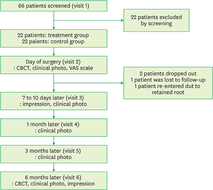
Figure 1 Flow diagram for the phases of the randomized controlled trial. CBCT: cone-beam computed tomography, VAS: visual analog scale.

Figure 2 (A) Polyworks superimposition of cast scans. Adjacent teeth were used as reference points. (B) A vector was projected from the designated 3D area. 3D: 3-dimensional.

Figure 3 (A) The sagittal image of CBCT from V2. (B) Calculation of height in the V6 image, which was cut similarly to the V2 image. CBCT: cone-beam computed tomography, V2: the day of surgery, V6: 6 months after the ridge preservation procedure.
Cited by 1 articles
-
A randomized controlled clinical study of periodontal tissue regeneration using an extracellular matrix-based resorbable membrane in combination with a collagenated bovine bone graft in intrabony defects
Sulhee Kim, Hyeyoon Chang, Jin wook Hwang, Sungtae Kim, Ki-Tae Koo, Tae-Il Kim, Yang-Jo Seol, Yong-Moo Lee, Young Ku, Jong-Ho Lee, In-Chul Rhyu
J Periodontal Implant Sci. 2017;47(6):363-371. doi: 10.5051/jpis.2017.47.6.363.
Reference
-
1. Amler MH, Johnson PL, Salman I. Histological and histochemical investigation of human alveolar socket healing in undisturbed extraction wounds. J Am Dent Assoc. 1960; 61:32–44.
Article2. Pietrokovski J, Massler M. Alveolar ridge resorption following tooth extraction. J Prosthet Dent. 1967; 17:21–27.
Article3. Araújo MG, Lindhe J. Dimensional ridge alterations following tooth extraction. An experimental study in the dog. J Clin Periodontol. 2005; 32:212–218.
Article4. Cardaropoli G, Araújo M, Lindhe J. Dynamics of bone tissue formation in tooth extraction sites. An experimental study in dogs. J Clin Periodontol. 2003; 30:809–818.5. Van der Weijden F, Dell’Acqua F, Slot DE. Alveolar bone dimensional changes of post-extraction sockets in humans: a systematic review. J Clin Periodontol. 2009; 36:1048–1058.
Article6. Schropp L, Wenzel A, Kostopoulos L, Karring T. Bone healing and soft tissue contour changes following single-tooth extraction: a clinical and radiographic 12-month prospective study. Int J Periodontics Restorative Dent. 2003; 23:313–323.7. Barone A, Aldini NN, Fini M, Giardino R, Calvo Guirado JL, Covani U. Xenograft versus extraction alone for ridge preservation after tooth removal: a clinical and histomorphometric study. J Periodontol. 2008; 79:1370–1377.
Article8. Iasella JM, Greenwell H, Miller RL, Hill M, Drisko C, Bohra AA, et al. Ridge preservation with freeze-dried bone allograft and a collagen membrane compared to extraction alone for implant site development: a clinical and histologic study in humans. J Periodontol. 2003; 74:990–999.
Article9. Vignoletti F, Matesanz P, Rodrigo D, Figuero E, Martin C, Sanz M. Surgical protocols for ridge preservation after tooth extraction. A systematic review. Clin Oral Implants Res. 2012; 23:Suppl 5. 22–38.
Article10. Araújo M, Linder E, Wennström J, Lindhe J. The influence of Bio-Oss Collagen on healing of an extraction socket: an experimental study in the dog. Int J Periodontics Restorative Dent. 2008; 28:123–135.11. Esposito M, Grusovin MG, Felice P, Karatzopoulos G, Worthington HV, Coulthard P. Interventions for replacing missing teeth: horizontal and vertical bone augmentation techniques for dental implant treatment. Cochrane Database Syst Rev. 2009; CD003607.
Article12. Hämmerle CH, Araújo MG, Simion M. Osteology Consensus Group 2011. Evidence-based knowledge on the biology and treatment of extraction sockets. Clin Oral Implants Res. 2012; 23:Suppl 5. 80–82.
Article13. Thalmair T, Fickl S, Schneider D, Hinze M, Wachtel H. Dimensional alterations of extraction sites after different alveolar ridge preservation techniques - a volumetric study. J Clin Periodontol. 2013; 40:721–727.
Article14. Lekovic V, Camargo PM, Klokkevold PR, Weinlaender M, Kenney EB, Dimitrijevic B, et al. Preservation of alveolar bone in extraction sockets using bioabsorbable membranes. J Periodontol. 1998; 69:1044–1049.
Article15. Bunyaratavej P, Wang HL. Collagen membranes: a review. J Periodontol. 2001; 72:215–229.
Article16. Zubery Y, Goldlust A, Alves A, Nir E. Ossification of a novel cross-linked porcine collagen barrier in guided bone regeneration in dogs. J Periodontol. 2007; 78:112–121.
Article17. Rothamel D, Benner M, Fienitz T, Happe A, Kreppel M, Nickenig HJ, et al. Biodegradation pattern and tissue integration of native and cross-linked porcine collagen soft tissue augmentation matrices - an experimental study in the rat. Head Face Med. 2014; 10:10.
Article18. Hwang JW, Kim S, Kim SW, Lee JH. Effect of extracellular matrix membrane on bone formation in a rabbit tibial defect model. Biomed Res Int. 2016; 2016:6715295.
Article19. Omran M, Min S, Abdelhamid A, Liu Y, Zadeh HH. Alveolar ridge dimensional changes following ridge preservation procedure: part-2 - CBCT 3D analysis in non-human primate model. Clin Oral Implants Res. 2016; 27:859–866.
Article20. Cardaropoli D, Tamagnone L, Roffredo A, Gaveglio L, Cardaropoli G. Socket preservation using bovine bone mineral and collagen membrane: a randomized controlled clinical trial with histologic analysis. Int J Periodontics Restorative Dent. 2012; 32:421–430.21. Jung RE, Philipp A, Annen BM, Signorelli L, Thoma DS, Hämmerle CH, et al. Radiographic evaluation of different techniques for ridge preservation after tooth extraction: a randomized controlled clinical trial. J Clin Periodontol. 2013; 40:90–98.
Article22. Fickl S, Zuhr O, Wachtel H, Stappert CF, Stein JM, Hürzeler MB. Dimensional changes of the alveolar ridge contour after different socket preservation techniques. J Clin Periodontol. 2008; 35:906–913.
Article
- Full Text Links
-
- Actions
-
Cited
- CITED
-
- Close
- Share
-
- Similar articles
-
- Improving oral rehabilitation through the preservation of the tissues through alveolar preservation
- Efficacy and safety of rhBMP/β-TCP in alveolar ridge preservation: a multicenter, randomized, open-label, comparative, investigator-blinded clinical trial
- The influence of membrane exposure on post-extraction dimensional change following ridge preservation technique
- A comparison between anorganic bone and collagen-preserving bone xenografts for alveolar ridge preservation: systematic review and future perspectives
- Compromised extraction sockets: a new classification and prevalence involving both soft and hard tissue loss
