Overexpression of Transforming Growth Factor-beta1 in the Valvular Fibrosis of Chronic Rheumatic Heart Disease
- Affiliations
-
- 1Department of Pathology, Inha University College of Medicine, Incheon, Korea. luciado@inha.ac.kr
- 2Department of Cardiovascular Surgery, Yonsei University College of Medicine, Seoul, Korea.
- 3Department of Pathology, Yonsei University College of Medicine, Seoul, Korea.
- 4Department of Internal Medicine, Ehwa University College of Medicine, Seoul, Korea.
- KMID: 1786843
- DOI: http://doi.org/10.3346/jkms.2008.23.1.41
Abstract
- For the purpose of determining the pathogenic role of transforming growth factor-beta1 (TGF-beta1) in the mechanism of chronic rheumatic heart disease, we evaluated the expression of TGF-beta1, proliferation of myofibroblasts, and changes in extracellular matrix components including collagen and proteoglycan in 30 rheumatic mitral valves and in 15 control valves. High TGF-beta1 expression was identified in 21 cases (70%) of rheumatic mitral valves, whereas only 3 cases (20%) of the control group showed high TGF-beta1 expression (p<0.001). Additionally, increased proliferation of myofibroblasts was observed in the rheumatic valves. High TGF-beta1 expression positively correlated with the proliferation of myofibroblasts (p=0.004), valvular fibrosis (p< 0.001), inflammatory cell infiltration (p=0.004), neovascularization (p=0.007), and calcification (p<0.001) in the valvular leaflets. The ratio of proteoglycan to collagen deposition inversely correlated with TGF-beta1 expression in mitral valves (p=0.040). In conclusion, an ongoing inflammatory process, the expression of TGF-beta1, and proliferation of myofibroblasts within the valves have a potential role in the valvular fibrosis, calcification, and changes in the extracellular matrix that lead to the scarring sequelae of rheumatic heart disease.
MeSH Terms
Figure
-
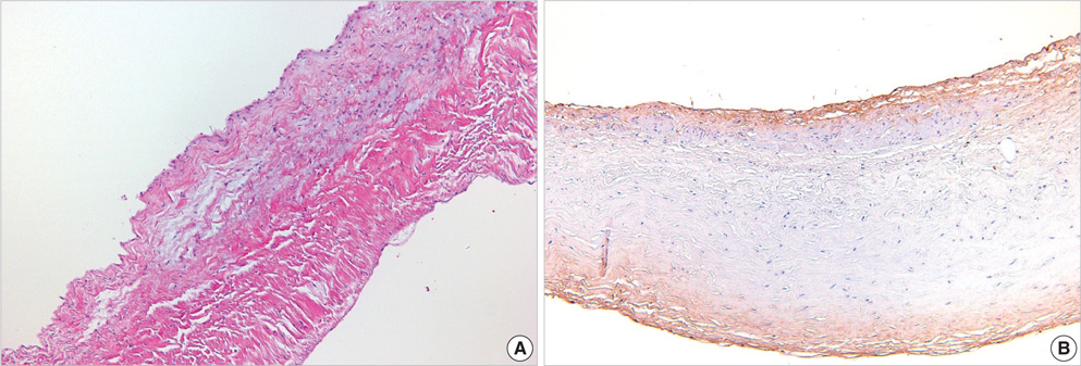
Fig. 1 Histologic and immunohistochemical findings of control valves. (A) Valvular leaflets obtained from the control group showed well-preserved leaflet architecture without fibrosis or inflammatory cell infiltration (H&E, ×40). (B) Immunohistochemical staining for TGF-β1 demonstrated positivity in the subendothelial stroma of valvular leaflets (TGF-β1, ×40).
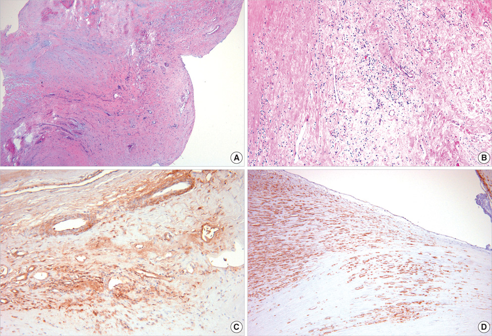
Fig. 2 Histologic and immunohistochemical findings of the rheumatic mitral valves. (A) Rheumatic mitral valves showed severe fibrosis and distorted architecture (H&E, ×40). (B) A high-power view demonstrated small thin-walled vessels and perivascular lymphocytic infiltration (H&E, ×200). (C) High TGF-β1 expression was seen in the endothelial cells and smooth muscle cells of the vessels, in the perivascular interstitial cells, and stroma of the valves (×200). (D) Myofibroblasts that were positive for SMA immunostaining were present in the subendothelial densely fibrotic area (×40).
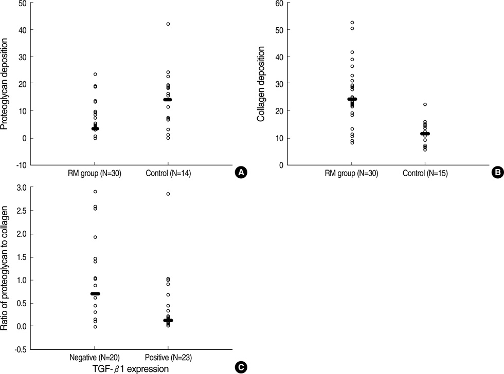
Fig. 3 Proteoglycan and collagen deposition in rheumatic mitral valves and control valves. (A) The proportion of proteoglycan deposited areas was lower in rheumatic mitral valve group (RM) than in the control group (black bars represented mean values: 3.75% vs. 13.2%) (p=0.014). (B) The rheumatic mitral valve group (RM) showed extensive collagen deposition compared to the control group (black bars represented mean values: 23% vs. 11.2%) (p<0.001). (C) The ratio of proteoglycan to collagen deposited areas was lower in the TGF-β1 positive group than in the TGF-β1 negative group (black bars represented mean values: 0.38 vs. 0.66) (p=0.040).
Reference
-
1. Schoen FJ. Cotran RS, Kumar V, Collins T, editors. The heart. Robbins pathologic basis of disease. 1999. 6th ed. Philadelphia: W.B. Saunders;543–599.
Article2. Rosai J. Rosai J, editor. Cardiovascular system. Ackerman's surgical pathology. 1996. 8th ed. St. Louis: Mosby;2173–2226.3. Veasy LG, Hill HR. Immunologic and clinical correlations in rheumatic fever and rheumatic heart disease. Pediatr Infect Dis J. 1997. 16:400–407.
Article4. Kemeny E, Grieve T, Marcus R, Sareli P, Zabriskie JB. Identification of mononuclear cells and T cell subsets in rheumatic valvulitis. Clin Immunol Immunopathol. 1989. 52:225–237.
Article5. Chen MC, Chang HW, Wu CJ, Yang CH, Yu TH, Chen CJ, Hung WC. Balance between plasma levels of tumor necrosis factor-alpha and interleukin-10 in rheumatic mitral stenosis. Cardiology. 2005. 104:171–175.6. Chiu-Braga YY, Hayashi SY, Schafranski M, Messias-Reason IJ. Further evidence of inflammation in chronic rheumatic valve disease (CRVD): high levels of advanced oxidation protein products (AOPP) and high sensitive C-reactive protein (hs-CRP). Int J Cardiol. 2006. 109:275–276.
Article7. Davutoglu V, Celik A, Aksoy M. Contribution of selected serum inflammatory mediators to the progression of chronic rheumatic valve disease, subsequent valve calcification and NYHA functional class. J Heart Valve Dis. 2005. 14:251–256.8. Bhatnagar A, grover A, ganguly NK. Superantigen-induced T cell responses in acute rheumatic fever and chronic rheumatic heart disease patients. Clin Exp Immunol. 1999. 116:100–106.
Article9. Lakos G, Melichian D, Wu M, Varga J. Increased bleomycin-induced skin fibrosis in mice lacking the Th1-specific transcription factor T-bet. Pathobiology. 2006. 73:224–237.
Article10. Kimura T, Ishii Y, Yoh K, Morishima Y, Iizuka T, Kiwamoto T, Matsuno Y, Homma S, Nomura A, Sakamoto T, Takahashi S, Sekizawa K. Overexpression of the transcription factor GATA-3 enhances the development of pulmonary fibrosis. Am J Pathol. 2006. 169:96–104.
Article11. Wen FQ, Liu X, Kobayashi T, Abe S, Fang Q, Kohyama T, Ertl R, Terasaki Y, Manouilova L, Rennard SI. Interferon-gamma inhibits transforming growth factor-beta production in human airway epithelial cells by targeting Smads. Am J Respir Cell Mol Biol. 2004. 30:816–822.12. Border WA, Noble NA. Transforming growth factor beta in tissue fibrosis. N Eng J Med. 1994. 331:1286–1292.13. O'Kane S, Ferguson MW. Transforming growth factor betas and wound healing. Int J Biochem Cell Biol. 1997. 29:63–78.14. Mutsaers SE, Bishop JE, McGrouther G, Laurent GJ. Mechanisms of tissue repair: from wound healing to fibrosis. Int J Biochem Cell Biol. 1997. 29:5–17.
Article15. Jian B, Connolly J, Savani RC, Narula N, Liang B, Levy R. Serotonin mechanisms in heart valve disease I: Serotonin-induced up-regulation of transforming growth factor-beta1 via G-protein signal transduction in aortic valve interstitial cells. Am J Pathol. 2002. 161:2111–2121.16. Jian B, Narula N, Li QY, Mohler ER 3rd, Levy RJ. Progression of aortic valve stenosis: TGF-β1 is present in calcified aortic valve cusps and promotes aortic valve interstitial cell calcification via apoptosis. Ann Thorac Surg. 2003. 75:457–465.
Article17. Chou HT, Chen CH, Tsai CH, Tsai FJ. Association between transforming growth factor-beta1 gene C-509T and T869C polymorphisms and rheumatic heart disease. Am Heart J. 2004. 148:181–186.18. Robiolio PA, Rigolin VH, Wilson JS, Harrison JK, Sanders LL, Bashore TM, Feldman JM. Carcinoid heart disease: Correlation of high serotonin levels with valvular abnormalities detected by cardiac catheterization and echocardiography. Circulation. 1995. 92:790–795.19. Ng CM, Cheng A, Myers LA, Martinez-Murillo F, Jie C, Bedja D, Gabrielson KL, Hausladen JM, Mecham RP, Judge DP, Dietz HC. TGFbeta-dependent pathogenesis of mitral valve prolapse in a mouse model of Marfan syndrome. J Clin Invest. 2004. 114:1586–1592.20. Ayoub EM, Toranta A, Bartley TD. Effect of valvular surgery on antibody to the group A streptococcal carbohydrate. Circulation. 1974. 50:144–150.
Article21. Golbasi Z, Ucar O, Keles T, Sahin A, Cagli K, Camsari A, Diker E, Aydogdu S. Increased levels of high sensitive C-reactive protein in patients with chronic rheumatic valve disease: evidence of ongoing inflammation. Eur J Heart Fail. 2002. 4:593–595.22. Guilherme L, Cury P, Demarchi LM, Coelho V, Abel L, Lopez AP, Oshiro SE, Aliotti S, Cunha-Neto E, Pomerantzeff PM, Tanaka AC, Kalil J. Rheumatic heart disease: proinflammatory cytokines play a role in the progression and maintenance of valvular lesions. Am J Pathol. 2004. 165:1583–1591.23. Gorelik L, Flavell RA. Transforming growth factor-beta in T-cell biology. Nat Rev Immunol. 2002. 2:46–53.24. Walker GA, Masters KS, Shah DN, Anseth KS, Leinwand LA. Valvular myofibroblast activation by transforming growth factor-beta: implications for pathological extracellular matrix remodeling in heart valve disease. Circ Res. 2004. 95:253–260.25. Lijnen P, Petrov V. Transforming growth factor-beta 1-induced collagen production in cultures of cardiac fibroblasts is the result of the appearance of myofibroblasts. Methods Find Exp Clin Pharmacol. 2002. 24:333–344.
Article26. Khan R, Sheppard R. Fibrosis in heart disease: understanding the role of transforming growth factor-β1 in cardiomyopathy, valvular disease and arrhythmia. Immunology. 2006. 118:10–24.27. Lis Y, Burleigh MC, Parker DJ, Child AH, Hogg J, Davies MJ. Biochemical characterization of individual normal, floppy and rheumatic human mitral valves. Biochem J. 1987. 244:597–603.
Article
- Full Text Links
-
- Actions
-
Cited
- CITED
-
- Close
- Share
-
- Similar articles
-
- Significant fibrosis after radiation therapy in a patient with Marfan syndrome
- TGF-beta-activated kinase-1: New insights into the mechanism of TGF-beta signaling and kidney disease
- Smads as therapeutic targets for chronic kidney disease
- Effects of TGF-beta1 Ribbon Antisense on CCl4-induced Liver Fibrosis
- Expression of Transforming Growth Factor-beta1 mRNA in Hepatocelluar Carcinoma and Surrounding Liver
