J Korean Med Sci.
2007 Jun;22(3):536-545. 10.3346/jkms.2007.22.3.536.
Quantitative Analysis of Aortic Atherosclerosis in Korean Female: A Necropsy Study
- Affiliations
-
- 1Department of Forensic Medicine, National Institute of Scientific Investigation, Seoul, Korea.
- 2Institute of Forensic Medicine, Pusan National University School of Medicine, Busan, Korea.
- 3Department of Anatomy, Sungkyunkwan University School of Medicine, Suwon, Korea. hdkim@med.skku.ac.kr
- KMID: 1778367
- DOI: http://doi.org/10.3346/jkms.2007.22.3.536
Abstract
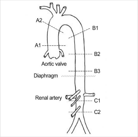
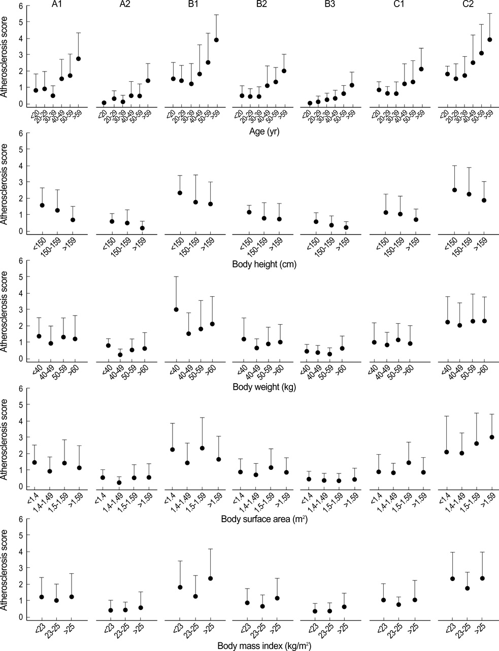
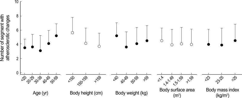
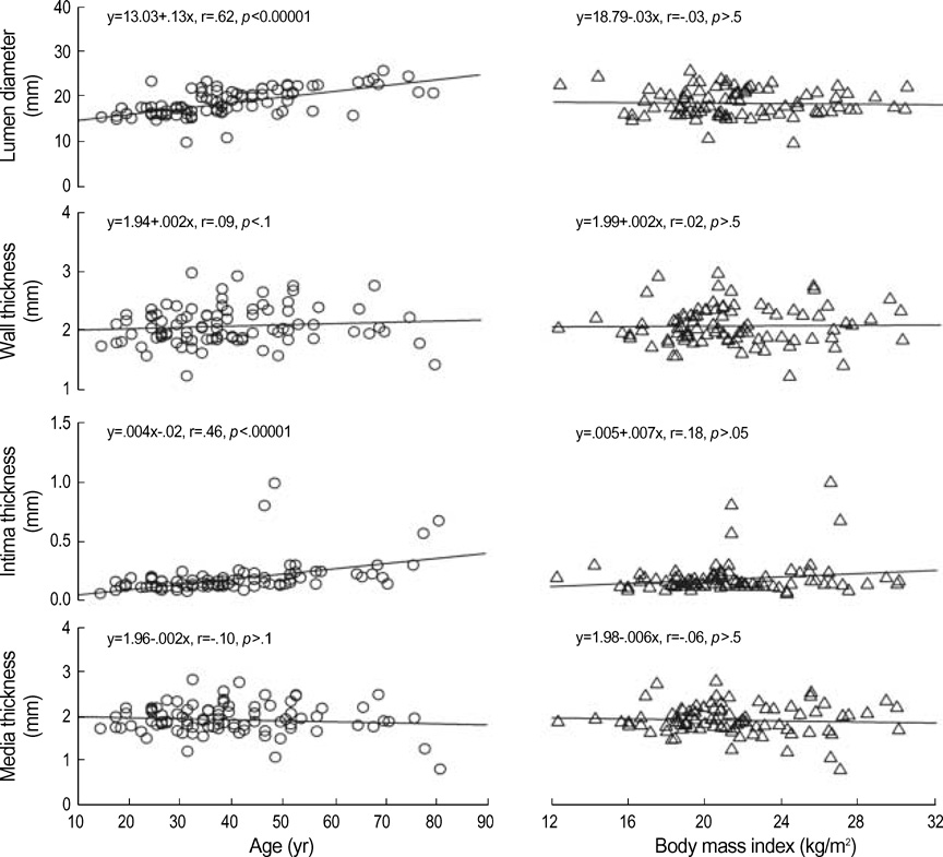
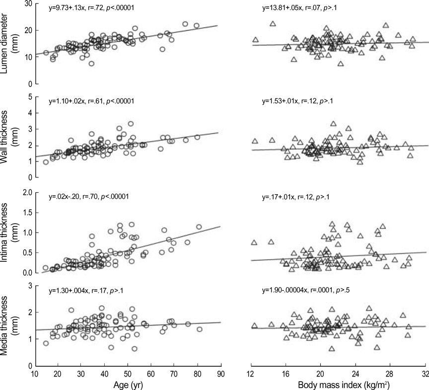
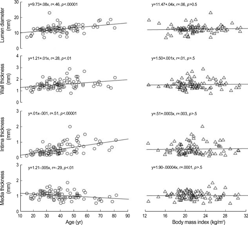
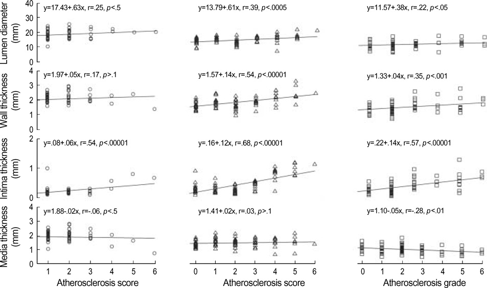
- To assess the regional difference and influence of the biological variables on atherosclerosis in female, we analyzed 7 segments of aorta (2 ascending, 3 thoracic, and 2 abdominal) from 90 superficially healthy Korean women (39+/-14 yr of age) who died from external causes. Tissue specimens were macroscopically examined and histopathologically divided into 7 grades for scoring (ATHERO, from 0=intact, to 6=thrombi formation). Lumen diameter (LD), wall thickness (WT), intima thickness (INT), and media thickness (MED) were obtained by computed morphometry. Atherosclerosis was common in the distal infrarenal (C2), proximal thoracic (B1), and proximal ascending (A1) segments. Total 95.6% of all subjects had atherosclerosis of variable degree in one or more segments, but an aneurysmal change was not found. The number of atherosclerotic segments and atherosclerosis score in the 7 segments increased with aging. However, the body size did not affect the aortic size and ATHERO. With aging, LD and INT of the A1, B1 and C2 increased (p<.00001); WT of the B1 and C2 increased (p<.01); and MED of C2 decreased (p<.01). LD and WT of the B1 and C2 (p<.05), INT of the A1, B1 and C2 (p<.00001) increased, and MED of C2 decreased (p<.01) with ATHERO. These data suggest that age is simple but a reliable parameter for estimating the progression of atherosclerosis.
Keyword
MeSH Terms
Figure
Reference
-
1. Johansen K, Kohler TR, Nicholls SC, Zieler RE, Clowes AW, Kazimers A. Ruptured aortic aneurysm: the Harborview experience. J Vasc Surg. 1991. 13:240–247.2. Brown PM, Zelt DT, Sobolev B. The risk of rupture in untreated aneurysms: the impact of size, gender, and expansion rate. J Vasc Surg. 2003. 37:280–284.
Article3. Newman AB, Arnold AM, Burke GL, O'Leary DH, Manolio TA. Cardiovascular disease and mortality in older adults with small abdominal aortic aneurysms detected by ultrasonography: the cardiovascular health study. Ann Intern Med. 2001. 134:182–190.
Article4. Bengtsson H, Bergqvist D, Sternby NH. Increasing prevalence of abdominal aortic aneurysms. A necropsy study. Eur J Surg. 1992. 158:19–23.5. Jousilahti P, Tuomilehto J, Vartiainen E, Pekkanen J, Puska P. Body weight, cardiovascular risk factors, and coronary mortality: 15-year follow-up of middle-aged men and women in eastern Finland. Circulation. 1996. 93:1372–1379.6. Duvall WL, Vorchheimer DA. Multi-bed vascular disease and atherothrombosis: scope of the problem. J Thromb Thrombolysis. 2004. 17:51–61.
Article7. Stern MP, Haffner SM. Body fat distribution and hyperinsulinemia as risk factors of diabetes and cardiovascular disease. Arteriosclerosis. 1986. 6:123–130.8. Calle EE, Thun MJ, Petrelli JM, Rodriguez C, Heath CW Jr. Body-mass index and mortality in a prospective cohort of U.S. adults. N Engl J Med. 1999. 34:1097–1105.
Article9. McGill HC Jr, McMahan CA, Herderick EE, Zieske AW, Malcom GT, Tracy RE, Strong JP. Obesity accelerates the progression of coronary atherosclerosis in young men. Circulation. 2002. 105:2712–2718.
Article10. Moon OR, Kim NS, Jang SM, Yoon TM, Kim SO. The relationship between body mass index and the prevalence of obesity-related diseases based on the 1995 National Health Interview Survey in Korea. Obes Rev. 2002. 3:191–196.
Article11. Stary HC, Chandler AB, Dinsmore RE, Fuster V, Glagov S, Insull W Jr, Rosenfeld ME, Schwartz CJ, Wagner WD, Wissler RW. A definition of advanced types of atherosclerotic lesions and a histological classification of atherosclerosis. A report from the committee on vascular lesions of the council on atherosclerosis, American Heart Association. Circulation. 1995. 92:1355–1374.12. Allison MA, Criqui Mh, Wright CM. Patterns and risk factors for systemic calcified atherosclerosis. Arterioscler Thromb Vasc Biol. 2004. 24:331–336.
Article13. Miwa Y, Tsushima M, Arima H, Kawano Y, Sasaguri T. Pulse pressure is an independent predictor for the progression of aortic wall calcification in patients with controlled hyperlipidemia. Hypertension. 2004. 43:536–540.
Article14. Liddington MI, Heather BP. The relationship between aortic diameter and body habitus. Eur J Vasc Surg. 1992. 6:89–92.
Article15. Sonneson B, Laenne T, Hansen F, Sandgren T. Infrarenal aortic diameter in healthy person. Eur J Vasc Surg. 1994. 8:89–95.16. Itani Y, Watanabe S, Masuda Y, Hanamura K, Asakura K, Sone S, Sunami Y, Miyamoto T. Measurement of aortic diameters and detection of asymptomatic aortic aneurysms in a mass screening program using a mobile helical computed tomography unit. Heart Vessels. 2002. 16:42–45.
Article17. Rexrode KM, Hennekens CH, Willett WC, Colditz GA, Stampfer MJ, Rich-Edwards JW, Speizer FE, Manson JE. A prospective study of body mass index, weight change, and risk of stroke in women. JAMA. 1997. 277:1539–1545.
Article18. Lakka TA, Lakka HM, Salonen R, Kaplan GA, Salonen JT. Abdomi nal obesity is associated with accelerated progression of carotid atherosclerosis in men. Atherosclerosis. 2001. 154:497–504.19. Schreyer SA, Lystig TC, Vick CM, LeBoeuf RC. Mice deficient in apolipoprotein E but not LDL receptors are resistant to accelerated atherosclerosis associated with obesity. Atherosclerosis. 2003. 171:49–55.
Article20. Wang J, Thornoton JC, Russel M, Burastero S, Heymsfield S, Pierson RN Jr. Asians have lower body mass index but higher percent body fat than do whites: comparison of anthropometric measurement. Am J Clin Nutr. 1994. 60:23–38.21. Ko GT, Chan JC, Cockram CS, Woo J. Prediction of hypertension, diabetes, dyslipidemia or albuminuria using simple anthropometric indexes in Hong Kong Chinese. Int J Obes Relat Metab Disord. 1999. 23:1136–1142.22. Nevill AM, Stewart AD, Olds R, Holt T. Relationship between adiposity and body size reveals limitations of BMI. Am J Phys Anthropol. 2006. 129:151–156.
Article23. Do KA, Green A, Guthrie JR, Dudley EC, Burger HG, Dennerstein L. Longitudinal study of risk factors for coronary heart disease across the menopausal transition. Am J Epidemiol. 2000. 151:584–593.
Article24. Zaydun G, Tomiyama H, Hashimoto H, Arai T, Koji Y, Yambe M, Motobe K, Hori S, Yamashina A. Menopause is an independent factor augmenting the age-related increase in arterial stiffness in the early postmenopausal phase. Atherosclerosis. 2006. 184:137–142.
Article25. Wanhainen A, Lundkvist J, Bergqvist D, Bjorck M. Cost-effectiveness of screening women for abdominal aortic aneurysm. J Vasc Surg. 2006. 43:908–914.
Article26. da Silva ES, Rodrigues AJ Jr, de Tolosa EM, Pereira PR, Zanoto A, Martins J. Variation of infrarenal aortic diameter: a necropsy study. J Vasc Surg. 1999. 29:920–927.
Article27. Fleischmann D, Hastie TJ, Dannegger FC, Paik DS, Tillsch M, Zarins CK, Rubin GD. Quantitative determination of age-related geometric changes in the normal abdominal aorta. J Vasc Surg. 2001. 33:97–105.
Article28. Zarins CK, Xu C, Glagov S. Atherosclerotic enlargement of the human abdominal aorta. Atherosclerosis. 2001. 155:157–164.
Article29. Zarins CK, Weisenberg E, Kolettis G, Stankunavicius R, Glagov S. Differential enlargement of artery segments in response to enlarging atherosclerotic plaques. J Vasc Surg. 1988. 7:386–394.
Article
- Full Text Links
- Actions
-
Cited
- CITED
-
- Close
- Share
- Similar articles
-
- Carotid Atherosclerosis as a Marker of Atherosclerosis of the Thoracic Aorta in the Elderly
- Transesophageal Echocardiographic Evaluation of Atherosclerosis
- Two Cases with Thoracic Aortic Dissection Combined with Fusiform Abdominal Aortic Aneurysm
- A Case of 51 Year Old Woman with Quadricuspid Aortic Valve Associated with Regurgitation
- Rupture of Ascending Thoracic Aortic Aneurysm in Postpartum: 2 Cases Report

