J Vet Sci.
2013 Dec;14(4):495-497. 10.4142/jvs.2013.14.4.495.
Percutaneous transplantation of human umbilical cord-derived mesenchymal stem cells in a dog suspected to have fibrocartilaginous embolic myelopathy
- Affiliations
-
- 1Department of Veterinary Surgery, College of Veterinary Medicine, Konkuk University, Seoul 143-701, Korea. hykim@konkuk.ac.kr
- 2Seoul Cord Blood Bank, Histostem Co., Seongnam 462-807, Korea.
- KMID: 1712319
- DOI: http://doi.org/10.4142/jvs.2013.14.4.495
Abstract
- The use of human umbilical cord blood-derived mesenchymal stem cells for cell transplantation therapy holds great promise for repairing spinal cord injury. Here we report the first clinical trial transplantation of human umbilical cord (hUCB)-derived mesenchymal stem cells (MSCs) into the spinal cord of a dog suspected to have fibrocartilaginous embolic myelopathy (FCEM) and that experienced a loss of deep pain sensation. Locomotor functions improved following transplantation in a dog. Based on our findings, we suggest that transplantation of hUCB-derived MSCs will have beneficial therapeutic effects on FCEM patients lacking deep pain sensation.
Keyword
MeSH Terms
Figure
-
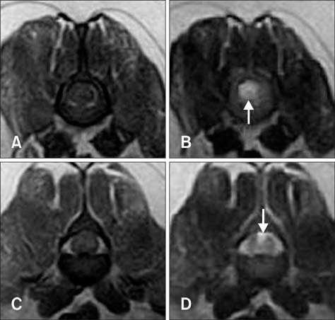
Fig. 1 T1-weighted (TR 530, TE 26) and T2-weighted (TR 3500, TE 90) MRI images of the dog 12 h (A~D) after the onset of clinical signs. The transverse T1-weighted image showed isointensity in the parenchyma of L2 (A) and L3 (C). Arrows: transverse T2-weighted image showing hyperintensity in the parenchyma of L2 (B) and L3 (D).
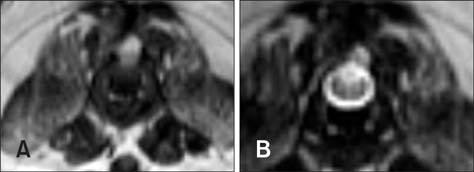
Fig. 2 T1-weighted (TR 550.0, TE 12.6) and T2-weighted (TR 4400, TE 96) MRI images of the dog 12 weeks after transplantation. The transverse T1-weighted image showed isointensity in the parenchyma of L3 (A). In the transverse T2-weighted image of L3, an area of hyperintensity was visible in the dorsal aspect of the spinal cord (B).
Reference
-
1. Abramson CJ, Garosi L, Platt SR, Dennis R, McConnell JF. Magnetic resonance imaging appearance of suspected ischemic myelopathy in dogs. Vet Radiol Ultrasound. 2005; 46:225–229.
Article2. Bagley RS. Fundamentals of Veterinary Clinical Neurology. 1st ed. Iowa: Blackwell;2005. p. 57–107.3. Cauzinille L, Kornegay JN. Fibrocartilaginous embolism of the spinal cord in dogs: review of 36 histologically confirmed cases and retrospective study of 26 suspected cases. J Vet Intern Med. 1996; 10:241–245.
Article4. Dasari VR, Spomar DG, Gondi CS, Sloffer CA, Saving KL, Gujrati M, Rao JS, Dinh DH. Axonal remyelination by cord blood stem cells after spinal cord injury. J Neurotrauma. 2007; 24:391–410.
Article5. De Risio L, Platt SR. Fibrocartilaginous embolic myelopathy in small animals. Vet Clin North Am Small Anim Pract. 2010; 40:859–869.
Article6. Fan CG, Zhang QJ, Tang FW, Han ZB, Wang GS, Han ZC. Human umbilical cord blood cells express neurotrophic factors. Neurosci Lett. 2005; 380:322–325.
Article7. Gandini G, Cizinauskas S, Lang J, Fatzer R, Jaggy A. Fibrocartilaginous embolism in 75 dogs: clinical findings and factors influencing the recovery rate. J Small Anim Pract. 2003; 44:76–80.
Article8. Grünenfelder FI, Weishaupt D, Green R, Steffen F. Magnetic resonance imaging findings in spinal cord infarction in three small breed dogs. Vet Radiol Ultrasound. 2005; 46:91–96.
Article9. Hawthorne JC, Wallace LJ, Fenner WR, Waters DJ. Fibrocartilaginous embolic myelopathy in miniature schnauzers. J Am Anim Hosp Assoc. 2001; 37:374–383.
Article10. Lee JH, Chang HS, Kang EH, Chung DJ, Choi CB, Lee JH, Hwang SH, Han H, Kim HY. Percutaneous transplantation of human umbilical cord blood-derived multipotent stem cells in a canine model of spinal cord injury. J Neurosurg Spine. 2009; 11:749–757.
Article11. Nakamoto Y, Ozawa T, Katakabe K, Nishiya K, Mashita T, Morita Y, Yasuda N, Ishii Y, Nakaichi M, Itamoto K. Usefulness of an early diagnosis for the favorable prognosis of fibrocartilaginous embolism diagnosed by magnetic resonance imaging in 10 small- to middle-sized dogs. Vet Res Commun. 2008; 32:609–617.
Article12. Nakamoto Y, Ozawa T, Katakabe K, Nishiya K, Yasuda N, Mashita T, Morita Y, Nakaichi M. Fibrocartilaginous embolism of the spinal cord diagnosed by characteristic clinical findings and magnetic resonance imaging in 26 dogs. J Vet Med Sci. 2009; 71:171–176.
Article13. Tarlov IM. Spinal cord compression studies. III. Time limits for recovery after gradual compression in dogs. AMA Arch Neurol Psychiatry. 1954; 71:588–597.
- Full Text Links
-
- Actions
-
Cited
- CITED
-
- Close
- Share
-
- Similar articles
-
- Differentiation of Osteoblast Progenitor Cells from Human Umbilical Cord Blood
- Difference in HLA-DR Expression of Human Umbilical Cord Blood Derived Mesenchymal Stem Cells after Tri-lineage Differentiation
- Therapy by traditional veterinary medicine in a case with canine fibrocartilaginous embolic myelopathy
- Stem Cell Transplantation in Umbilical Cord Blood(I) Expansion Effects of Stem Cells in Umbilical Cord Blood with Various Hematopoietic Growth Factors
- Endothelial progenitor cells and mesenchymal stem cells from human cord blood
