Sentinel Node Mapping of VX2 Carcinoma in Rabbit Thigh with CT Lymphography Using Ethiodized Oil
- Affiliations
-
- 1Department of Radiology, Seoul National University Bundang Hospital, Seongnam 463-707, Korea. yhkrad@gmail.com
- 2Department of Pathology, Seoul National University Bundang Hospital, Seongnam 463-707, Korea.
- 3Department of Radiology, Seoul National University Hospital, Seoul National University College of Medicine, Seoul 110-744, Korea.
- 4Department of Radiology, Gangnam Severance Hospital, Yonsei University College of Medicine, Seoul 135-720, Korea.
- KMID: 1711474
- DOI: http://doi.org/10.3348/kjr.2014.15.1.29
Abstract
OBJECTIVE
To assess the feasibility of computed tomography (CT) lymphography using ethiodized oil for sentinel node mapping in experimentally induced VX2 carcinoma in the rabbit thigh.
MATERIALS AND METHODS
This experiment received approval from the institutional animal use and care administrative advisory committee. Twenty-three rabbits with VX2 carcinoma in the thigh underwent CT before and after (1 hour, 2 hour) peritumoral injection of 2 mL ethiodized oil. After the CT examination, sentinel nodes were identified by peritumoral injection of methylene blue and subsequently removed. The retrieved sentinel and non-sentinel lymph nodes were investigated with radiographic and pathologic examinations. Based on the comparison of CT findings with those of radiographic and pathologic examinations, the diagnostic performance of CT for sentinel node identification was assessed.
RESULTS
All 23 rabbits showed 53 ethiodized oil retention nodes on post-injection CT and specimen radiography, and 52 methylene blue-stained nodes at the right femoroiliac area. Of the 52 blue-stained sentinel nodes, 50 nodes demonstrated ethiodized oil retention. Thus, the sentinel node detection rate of CT was 96% (50 of 52). On pathologic examination, 28 sentinel nodes in 17 rabbits (nodes/rabbit, mean +/- standard deviation, 1.7 +/- 0.6) harbored metastasis. Twenty seven of the 28 metastatic sentinel nodes were found to have ethiodized oil retention.
CONCLUSION
Computed tomography lymphography using ethiodized oil may be feasible for sentinel node mapping in experimentally induced VX2 carcinoma in the rabbit thigh.
MeSH Terms
-
Animals
Carcinoma/pathology/*radiography/secondary
Contrast Media/*diagnostic use
Ethiodized Oil/*diagnostic use
Feasibility Studies
Female
Injections
Lymph Node Excision/methods
Lymph Nodes/pathology/*radiography
Lymphatic Metastasis/radiography
Lymphography/*methods
Rabbits
Thigh
Tomography, X-Ray Computed/*methods
Contrast Media
Ethiodized Oil
Figure
-

Fig. 1 CT lymphography after ethiodized oil injection at peritumoral area in rabbit thigh. A, B. Noncontrast CT images obtained 1 (A) and 2 (B) hours after ethiodized oil injection show nodular high attenuation areas (arrows), suggesting ethiodized oil retention in right femoroiliac lymph nodes. C. Photography obtained after resection of blue-stained lymphatic basin shows blue-stained lymph nodes (arrows). D. Specimen radiography shows ethiodized oil retention (arrows) in blue-stained lymph nodes. E. Photomicrography of removed node demonstrates intranodal lipid droplets (red droplets, arrows), indicating intra-nodal ethiodized oil retention (Oil-Red-O stain; original magnification, × 40).
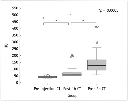
Fig. 2 Box-and-whisker plots of measured nodal attenuation values in Hounsfield unit (HU) at noncontrast CT before (pre-injection CT), 1 hour (post-1h CT) and 2 hours (post-2h CT) after ethiodized oil injection. Differences of attenuation values between all three pairs of pre- and post-1h and post-2h CT images were statistically significant (p < 0.0005, respectively).
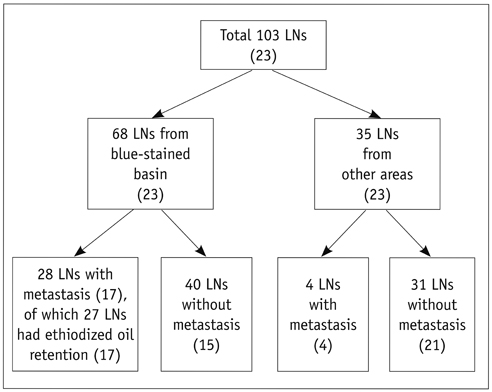
Fig. 3 Flow diagram of histologic findings of retrieved lymph nodes. Numbers in parentheses are numbers of rabbits. LN = lymph node
Reference
-
1. Morton DL, Wen DR, Wong JH, Economou JS, Cagle LA, Storm FK, et al. Technical details of intraoperative lymphatic mapping for early stage melanoma. Arch Surg. 1992; 127:392–399.2. Veronesi U, Paganelli G, Galimberti V, Viale G, Zurrida S, Bedoni M, et al. Sentinel-node biopsy to avoid axillary dissection in breast cancer with clinically negative lymph-nodes. Lancet. 1997; 349:1864–1867.3. Borgstein PJ, Pijpers R, Comans EF, van Diest PJ, Boom RP, Meijer S. Sentinel lymph node biopsy in breast cancer: guidelines and pitfalls of lymphoscintigraphy and gamma probe detection. J Am Coll Surg. 1998; 186:275–283.4. De Cicco C, Cremonesi M, Luini A, Bartolomei M, Grana C, Prisco G, et al. Lymphoscintigraphy and radioguided biopsy of the sentinel axillary node in breast cancer. J Nucl Med. 1998; 39:2080–2084.5. Linehan DC, Hill AD, Akhurst T, Yeung H, Yeh SD, Tran KN, et al. Intradermal radiocolloid and intraparenchymal blue dye injection optimize sentinel node identification in breast cancer patients. Ann Surg Oncol. 1999; 6:450–454.6. Morton DL, Thompson JF, Cochran AJ, Mozzillo N, Elashoff R, Essner R, et al. Sentinel-node biopsy or nodal observation in melanoma. N Engl J Med. 2006; 355:1307–1317.7. Park DJ, Lee HJ, Lee HS, Kim WH, Kim HH, Lee KU, et al. Sentinel node biopsy for cT1 and cT2a gastric cancer. Eur J Surg Oncol. 2006; 32:48–54.8. Suga K, Ogasawara N, Okada M, Matsunaga N. Interstitial CT lymphography-guided localization of breast sentinel lymph node: preliminary results. Surgery. 2003; 133:170–179.9. Minato M, Hirose C, Sasa M, Nishitani H, Hirose Y, Morimoto T. 3-dimensional computed tomography lymphography-guided identification of sentinel lymph nodes in breast cancer patients using subcutaneous injection of nonionic contrast medium: a clinical trial. J Comput Assist Tomogr. 2004; 28:46–51.10. Suga K, Yuan Y, Okada M, Matsunaga N, Tangoku A, Yamamoto S, et al. Breast sentinel lymph node mapping at CT lymphography with iopamidol: preliminary experience. Radiology. 2004; 230:543–552.11. Tangoku A, Yamamoto S, Suga K, Ueda K, Nagashima Y, Hida M, et al. Sentinel lymph node biopsy using computed tomography-lymphography in patients with breast cancer. Surgery. 2004; 135:258–265.12. Suga K, Shimizu K, Kawakami Y, Tangoku A, Zaki M, Matsunaga N, et al. Lymphatic drainage from esophagogastric tract: feasibility of endoscopic CT lymphography for direct visualization of pathways. Radiology. 2005; 237:952–960.13. Suga K, Yamamoto S, Tangoku A, Oka M, Kawakami Y, Matsunaga N. Breast sentinel lymph node navigation with three-dimensional interstitial multidetector-row computed tomographic lymphography. Invest Radiol. 2005; 40:336–342.14. Kim YH, Lee YJ, Park JH, Lee KH, Lee HS, Park YS, et al. Early gastric cancer: feasibility of CT lymphography with ethiodized oil for sentinel node mapping. Radiology. 2013; 267:414–421.15. Handal JA, Schulz JF, Florez GB, Kwok SC, Khurana JS, Samuel SP. Creation of rabbit bone and soft tissue tumor using cultured VX2 cells. J Surg Res. 2013; 179:e127–e132.16. Lei Z, Ma H, Xu N, Xi H. The evaluation of anti-angiogenic treatment effects for implanted rabbit VX2 breast tumors using functional multi-slice spiral computed tomography (f-MSCT). Eur J Radiol. 2011; 78:277–281.17. Kim YH, Choi BI, Cho WH, Lim S, Moon WK, Han JK, et al. Dynamic contrast-enhanced MR imaging of VX2 carcinomas after X-irradiation in rabbits: comparison of gadopentetate dimeglumine and a macromolecular contrast agent. Invest Radiol. 2003; 38:539–549.18. Sutton R, Tsopelas C, Kollias J, Chatterton BE, Coventry BJ. Sentinel node biopsy and lymphoscintigraphy with a technetium 99m labeled blue dye in a rabbit model. Surgery. 2002; 131:44–49.19. Wu H, Ying H, Xi X, Shen N, Shu Y, Hoffman MR, et al. Localization of the sentinel lymph node in tongue VX2 carcinoma via indirect CT lymphography combined with methylene blue dye injection. Acta Otolaryngol. 2010; 130:503–510.20. Daigeler A, Kuhnen C, Moritz R, Stricker I, Goertz O, Tilkorn D, et al. Lymph node metastases in soft tissue sarcomas: a single center analysis of 1,597 patients. Langenbecks Arch Surg. 2009; 394:321–329.21. Fong Y, Coit DG, Woodruff JM, Brennan MF. Lymph node metastasis from soft tissue sarcoma in adults. Analysis of data from a prospective database of 1772 sarcoma patients. Ann Surg. 1993; 217:72–77.22. Behranwala KA, A'Hern R, Omar AM, Thomas JM. Prognosis of lymph node metastasis in soft tissue sarcoma. Ann Surg Oncol. 2004; 11:714–719.23. Sawamura C, Matsumoto S, Shimoji T, Ae K, Okawa A. Lymphadenectomy and histologic subtype affect overall survival of soft tissue sarcoma patients with nodal metastases. Clin Orthop Relat Res. 2013; 471:926–931.24. Andreou D, Boldt H, Werner M, Hamann C, Pink D, Tunn PU. Sentinel node biopsy in soft tissue sarcoma subtypes with a high propensity for regional lymphatic spread--results of a large prospective trial. Ann Oncol. 2013; 24:1400–1405.25. Giuliano AE, Haigh PI, Brennan MB, Hansen NM, Kelley MC, Ye W, et al. Prospective observational study of sentinel lymphadenectomy without further axillary dissection in patients with sentinel node-negative breast cancer. J Clin Oncol. 2000; 18:2553–2559.26. Feldman SM, Krag DN, McNally RK, Moor BB, Weaver DL, Klein P. Limitation in gamma probe localization of the sentinel node in breast cancer patients with large excisional biopsy. J Am Coll Surg. 1999; 188:248–254.27. Dupont E, Cox C, Shivers S, Salud C, Nguyen K, Cantor A, et al. Learning curves and breast cancer lymphatic mapping: institutional volume index. J Surg Res. 2001; 97:92–96.28. Mariani G, Moresco L, Viale G, Villa G, Bagnasco M, Canavese G, et al. Radioguided sentinel lymph node biopsy in breast cancer surgery. J Nucl Med. 2001; 42:1198–1215.29. Tangoku A, Seike J, Nakano K, Nagao T, Honda J, Yoshida T, et al. Current status of sentinel lymph node navigation surgery in breast and gastrointestinal tract. J Med Invest. 2007; 54:1–18.30. Schmid-Schönbein GW. Microlymphatics and lymph flow. Physiol Rev. 1990; 70:987–1028.31. Ikomi F, Hanna GK, Schmid-Schönbein GW. Mechanism of colloidal particle uptake into the lymphatic system: basic study with percutaneous lymphography. Radiology. 1995; 196:107–113.32. Kan Z, McCuskey PA, Wright KC, Wallace S. Role of Kupffer cells in iodized oil embolization. Invest Radiol. 1994; 29:990–993.33. Davidson JW, Fletch AL, McIlmoyle G, Roeck W. The technique and applications of lymphography. Can J Comp Med. 1973; 37:130–138.34. Guermazi A, Brice P, Hennequin C, Sarfati E. Lymphography: an old technique retains its usefulness. Radiographics. 2003; 23:1541–1558. discussion 1559-1560.35. Tanaka Y, Sawada S, Yoshida M, Murata T. Clinical evaluation of computed tomography images in esophageal cancer after submucosal injection of iodized oil. J Comput Tomogr. 1985; 9:125–132.36. Karanov SI. Endoscopic indirect lymphography in gastric cancer. Endoscopy. 1988; 20:241–243.37. Dietrich A, Wuttke M, Walter F, De Leon I, Kloeppel R, Schoenfelder M. Indirect lymph node lymphangiography using an iodine-based contrast medium and projection radiography following submucosal injection in a rabbit model. Langenbecks Arch Surg. 2002; 387:315–319.38. Kazem I, Nedwich A, Mortel R, Honda T. Comparative histological changes in the normal lymph node following ethiodol lymphography and colloidal gold-198 lymphscanning. Clin Radiol. 1971; 22:382–388.39. Clément O, Luciani A. Imaging the lymphatic system: possibilities and clinical applications. Eur Radiol. 2004; 14:1498–1507.40. Chung YE, Hyung WJ, Kweon S, Lim SJ, Choi J, Lee MH, et al. Feasibility of interstitial CT lymphography using optimized iodized oil emulsion in rats. Invest Radiol. 2010; 45:142–148.
- Full Text Links
-
- Actions
-
Cited
- CITED
-
- Close
- Share
-
- Similar articles
-
- Local tissue reaction after injection of contrast media on gastric wall of mouse: experimental study for application of contrast media to computed tomography lymphography
- Sentinel node mapping in endometrial cancer
- Preoperative Imaging of Sentinel Lymph Nodes in Gastric Cancer Using CT Lymphography
- A critical assessment on the role of sentinel node mapping in endometrial cancer
- Profound Hypotension after an Intradermal Injection of Indigo Carmine for Sentinel Node Mapping
