Tuberc Respir Dis.
2008 Mar;64(3):236-239. 10.4046/trd.2008.64.3.236.
A Pulmonary Sarcoidosis Manifesting as a Rare Atypical Pattern and Distribution
- Affiliations
-
- 1Department of Radiology, Korea University Guro Hospital, Korea University College of Medicine, Seoul, Korea. keyrad@korea.ac.kr
- 2Department of Pathology, Korea University Guro Hospital, Korea University College of Medicine, Seoul, Korea.
- 3Department of Internal Medicine, Korea University Guro Hospital, Korea University College of Medicine, Seoul, Korea.
- KMID: 1478188
- DOI: http://doi.org/10.4046/trd.2008.64.3.236
Abstract
- A unique case of atypical pulmonary sarcoidosis in a 62-year-old man complaining of dyspnea is presented. Chest CT scan showed an unusual pattern and distribution of pulmonary sarcoidosis manifesting mainly as reticular densities, interlobular septal thickening, and ground-glass opacities, in the subpleural and lower lung predominancy. However, a surgical lung biopsy revealed classical findings of sarcoidosis. Knowledge of this atypical pulmonary involvement may improve understanding sarcoidosis as the great masquerader.
Keyword
Figure
-
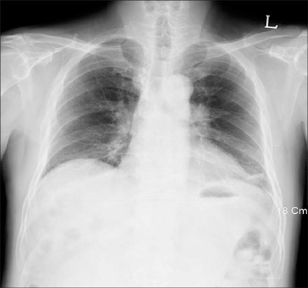
Figure 1 Chest PA view shows bilateral hilar lymphadenopathies.
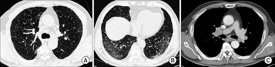
Figure 2 Thin-section CT with lung window setting (A, B) shows fine reticular densities, faint ground-glass opacities, and interlobular septal thickenings on both lungs, predominantly the lower subpleural lung zone. Enhanced chest CT scan with a mediastinal window setting (C) shows bilateral mediastinal and hilar lymphadenopathies.
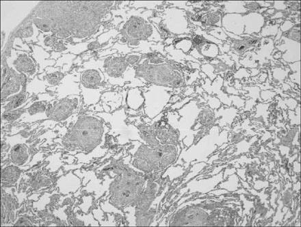
Figure 3 Surgical lung biopsy specimen shows extensive infiltration by noncaseating granulomas distributed along the pleura, interlobular septa, and bronchovascular bundles (H&E stain, ×40).
Reference
-
1. Nunes H, Brillet PY, Valeyre D, Brauner MW, Wells AU. Imaging in sarcoidosis. Semin Respir Crit Care Med. 2007. 28:102–120.2. Vagal AS, Shipley R, Meyer CA. Radiological manifestations of sarcoidosis. Clin Dermatol. 2007. 25:312–325.3. Koyama T, Ueda H, Togashi K, Umeoka S, Kataoka M, Nagai S. Radiologic manifestations of sarcoidosis in various organs. Radiographics. 2004. 24:87–104.4. Hennebicque AS, Nunes H, Brillet PY, Moulahi H, Valeyre D, Brauner MW. CT findings in severe thoracic sarcoidosis. Eur Radiol. 2005. 15:23–30.5. Hamper UM, Fishman EK, Khouri NF, Johns CJ, Wang KP, Siegelman SS. Typical and atypical CT manifestations of pulmonary sarcoidosis. J Comput Assist Tomogr. 1986. 10:928–936.6. Müller NL, Kullnig P, Miller RR. The CT findings of pulmonary sarcoidosis: analysis of 25 patients. AJR Am J Roentgenol. 1989. 152:1179–1182.7. Nishimura K, Itoh H, Kitaichi M, Nagai S, Izumi T. Pulmonary sarcoidosis: correlation of CT and histopathologic findings. Radiology. 1993. 189:105–109.8. Honda O, Johkoh T, Ichikado K, Yoshida S, Mihara N, Higashi M, et al. Comparison of high resolution CT findings of sarcoidosis, lymphoma, and lymphangitic carcinoma: is there any difference of involved interstitium? J Comput Assist Tomogr. 1999. 23:374–379.9. Johkoh T, Müller NL, Pickford HA, Hartman TE, Ichikado K, Akira M, et al. Lymphocytic interstitial pneumonia: thin-section CT findings in 22 patients. Radiology. 1999. 212:567–572.
- Full Text Links
-
- Actions
-
Cited
- CITED
-
- Close
- Share
-
- Similar articles
-
- Subcutaneous Sarcoidosis of the Distal Lower Leg in a Middle-Aged Woman Associated with Pulmonary Sarcoidosis: a Case Report
- Gastric Involvement of Pulmonary Sarcoidosis
- A Case of Pulmonary Sarcoidosis in a 6-year-old Girl
- Typical and Atypical Manifestations of Intrathoracic Sarcoidosis
- A case of sarcoidosis associated with acute tubular necrosis and pulmonary hemorrhage
