Korean J Orthod.
2010 Jun;40(3):134-144. 10.4041/kjod.2010.40.3.134.
Three dimensional analysis of the upper airway and facial morphology in children with Class II malocclusion using cone-beam computed tomography
- Affiliations
-
- 1Department of Orthodontics, Kangdong Sacred Heart Hospital, Hallym University Medical Center, Korea. dentpark64@hanmail.net
- 2Graduate School of Hallym University, Korea.
- 3Public Health Center of Namyangju-si, Korea.
- KMID: 1459567
- DOI: http://doi.org/10.4041/kjod.2010.40.3.134
Abstract
OBJECTIVE
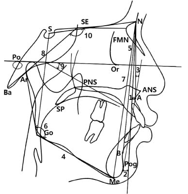
The aim of this study was to evaluate the volumes and areas of the upper airways in children with Class II malocclusion, using three dimensional cone-beam computed tomography (CBCT) and to compare the volumetric and cross-sectional measurements and cephalometric variables to investigate possible relationships between the upper airway and facial morphology.
METHODS
CBCT scans were obtained from 37 subjects (17 boys and 20 girls; average age, 11.02 years). The upper airway volumes and areas were measured, and compared with cephalometric variables.
RESULTS
The area of the PNS-posterior plane (SPP) was significantly smaller in the Class II malocclusion group (p < 0.05). Also, the volumetric and cross-sectional measurements were lower in Class II than in Class I malocclusion groups, although the differences were not significant between the two groups (p > 0.05). The Class II malocclusion group showed significantly smaller values of PFH, mandibular body length, pog to N perp and showed larger values of FMA, ANB, and facial convexity than the Class I malocclusion group. The volume of the upper airway in front of PNS point (WN) showed negative correlation with ANB (p < 0.05).
CONCLUSIONS
The Class II malocclusion group had a narrower upper airway associated with a decreased posterior facial height and a divergent growth pattern than the Class I malocclusion group.
Keyword
Figure
Reference
-
1. Ceylan I, Oktay H. A study on the pharyngeal size in different skeletal patterns. Am J Orthod Dentofacial Orthop. 1995. 108:69–75.
Article2. Holmberg H, Linder-Aronson S. Cephalometric radiographs as a means of evaluating the capacity of the nasal and nasopharyngeal airway. Am J Orthod. 1979. 76:479–490.
Article3. Joseph AA, Elbaum J, Cisneros GJ, Eisig SB. A cephalometric comparative study of the soft tissue airway dimensions in persons with hyperdivergent and normodivergent facial patterns. J Oral Maxillofac Surg. 1998. 56:135–139.
Article4. Ricketts RM. Respiratory obstruction syndrome. Am J Orthod. 1968. 54:495–507.5. Lopatiene K, Babarskas A. Malocclusion and upper airway obstruction. Medicina (Kaunas). 2002. 38:277–283.6. Angle E. Treatment of malocclusion of the teeth. 1907. 7th ed. Philadelphia: SS White Manufacturing Company;45.7. Linder-Aronson S. Adenoids. Their effect on mode of breathing and nasal airflow and their relationship to characteristics of the facial skeleton and the denition. A biometric, rhino-manometric and cephalometro-radiographic study on children with and without adenoids. Acta Otolaryngol Suppl. 1970. 265:1–132.8. Moore A. Observations on mouth breathing. Bull N Z Soc Periodontol. 1972. 33:9–11.9. Hwang CJ, Ryu YK. A longitudinal study of nasopharynx and adenoid growth of Korean children. Korean J Orthod. 1985. 15:93–104.10. Lee YS, Kim JC. A cephalometric study on the airway size according to the types of the malocclusion. Korean J Orthod. 1995. 25:19–29.11. Lee YS, Baik HS, Lee KJ, Yu HS. The structural change in the hyoid bone and upper airway after orthognathic surgery for skeletal class III anterior open bite patients using 3-dimensional computed tomography. Korean J Orthod. 2009. 39:72–82.
Article12. Alves PV, Zhao L, O'Gara M, Patel PK, Bolognese AM. Three-dimensional cephalometric study of upper airway space in skeletal class II and III healthy patients. J Craniofac Surg. 2008. 19:1497–1507.
Article13. Chang HS, Baik HS. A proposal of landmarks for craniofacial analysis using three-dimensional CT imaging. Korean J Orthod. 2002. 32:313–325.14. Iwasaki T, Hayasaki H, Takemoto Y, Kanomi R, Yamasaki Y. Oropharyngeal airway in children with Class III malocclusion evaluated by cone-beam computed tomography. Am J Orthod Dentofacial Orthop. 2009. 136:318.e1–318.e9.
Article15. Kim YI, Kim SS, Son WS, Park SB. Pharyngeal airway analysis of different craniofacial morphology using cone-beam computed tomography (CBCT). Korean J Orthod. 2009. 39:136–145.
Article16. Dahlberg G. Statistical methods for medical and biological students. 1940. London: G. Allen & Unwin Ltd;1–140.17. Lagravère MO, Carey J, Toogood RW, Major PW. Three-dimensional accuracy of measurements made with software on cone-beam computed tomography images. Am J Orthod Dentofacial Orthop. 2008. 134:112–116.
Article18. Major MP, Flores-Mir C, Major PW. Assessment of lateral cephalometric diagnosis of adenoid hypertrophy and posterior upper airway obstruction: a systematic review. Am J Orthod Dentofacial Orthop. 2006. 130:700–708.
Article19. Abramson ZR, Susarla S, Tagoni JR, Kaban L. Three-dimensional computed tomographic analysis of airway anatomy. J Oral Maxillofac Surg. 2010. 68:363–371.
Article20. Fouke JM, Strohl KP. Effect of position and lung volume on upper airway geometry. J Appl Physiol. 1987. 63:375–380.
Article21. Pae EK, Lowe AA, Sasaki K, Price C, Tsuchiya M, Fleetham JA. A cephalometric and electromyographic study of upper airway structures in the upright and supine positions. Am J Orthod Dentofacial Orthop. 1994. 106:52–59.
Article22. Proffit WR. Proffit WR, Fields HW, Sarver DM, editors. Concepts of growth and development. Contemporary orthodontics. 2007. 4th ed. St Louis: Mosby;27–29.23. Hwang YI, Lee KH, Lee KJ, Kim SC, Cho HJ, Cheon SH, et al. Effect of airway and tongue in facial morphology of prepubertal Class I, II children. Korean J Orthod. 2008. 38:74–82.
Article24. McNamara JA. Influence of respiratory pattern on craniofacial growth. Angle Orthod. 1981. 51:269–300.
Article25. Subtelny JD, Baker HK. The significance of adenoid tissue in velopharyngeal function. Plast Reconstr Surg. 1956. 17:235–250.
Article26. Mergen DC, Jacobs RM. The size of nasopharynx associated with normal occlusion and Class II malocclusion. Angle Orthod. 1970. 40:342–346.27. Kim YJ, Bok GS, Lee KH, Hwang YI, Park YH. The relationship between upper airway width and facial growth changes in orthodontic treatment of growing children. Korean J Orthod. 2009. 39:168–176.
Article28. Behlfelt K, Linder-Aronson S, McWilliam J, Neander P, Laage-Hellman J. Cranio-facial morphology in children with and without enlarged tonsils. Eur J Orthod. 1990. 12:233–243.
Article29. Solow B, Siersbaek-Nielsen S, Greve E. Airway adequacy, head posture, and craniofacial morphology. Am J Orthod. 1984. 86:214–223.
Article30. Sosa FA, Graber TM, Muller TP. Postpharyngeal lymphoid tissue in Angle Class I and Class II malocclusions. Am J Orthod. 1982. 81:299–309.
Article
- Full Text Links
- Actions
-
Cited
- CITED
-
- Close
- Share
- Similar articles
-
- Three dimensional cone-beam CT study of upper airway change after mandibular setback surgery for skeletal Class III malocclusion patients
- Position of the hyoid bone and its correlation with airway dimensions in different classes of skeletal malocclusion using cone-beam computed tomography
- A Study on the Nasal Index of Malocclusion Patients Using Cone-Beam Computed Tomography 3D Program
- Cone-beam computed tomographic evaluation of the temporomandibular joint and dental characteristics of patients with Class II subdivision malocclusion and asymmetry
- Pharyngeal airway analysis of different craniofacial morphology using cone-beam computed tomography (CBCT)

