Immunohistochemical localization of cyclic AMP-responsive element binding protein (CREB)-binding protein in the pig retina during postnatal development
- Affiliations
-
- 1Department of Veterinary Anatomy, Veterinary Medical Research Institute, College of Veterinary Medicine, Jeju National University, Jeju, Korea. shint@jejunu.ac.kr
- 2Department of Anatomy, Graduate School of Medicine, Jeju National University, Jeju, Korea.
- 3Department of Veterinary Anatomy, College of Veterinary Medicine, Chonnam National University, Gwangju, Korea. moonc@chonnam.ac.kr
- KMID: 1447424
- DOI: http://doi.org/10.5115/acb.2011.44.2.143
Abstract
- This study evaluated the cellular localization of cyclic AMP-responsive element binding protein-binding protein (CBP) expression in pig retinas during postnatal development. Immunohistochemistry and Western blot analysis were performed on retinal tissue from 2-day-old, 5-week-old, and 6-month-old pigs. Western blot analysis detected the expression of CBP in the retinas of 2-day-old piglets and showed that it was significantly decreased in the retinas of 5-week-old and 6-month-old pigs. Immunohistochemically, CBP was intensely immunostained in protein kinase C alpha (PKCalpha)-positive-bipolar cells, glutamine synthetase-positive Muller cells, and in ganglion cells in 2-day-old piglets. CBP was detected weakly in the inner plexiform, outer nuclear, and rod and cone layers. CBP immunoreactivity in the ganglion cell layer was decreased in the retinas of 5-week-old and 6-month-old pigs, while clear CBP expression detected in the neurite of PKCalpha-positive bipolar cells in the inner nuclear layer. In addition, CBP immunoreactivity in Muller cells and glial fibrillary acidic protein-positive glial processes was particularly noteworthy in pig retinas, but not in rat retinas. The results indicate that CBP is expressed differentially in the retinal neurons and glial cells according to growth and animal species, and may play an important role in homeostasis in Muller cells, neurite extention in bipolar cells, and signal transduction in photoreceptor cells in the porcine retina.
Keyword
MeSH Terms
Figure
-
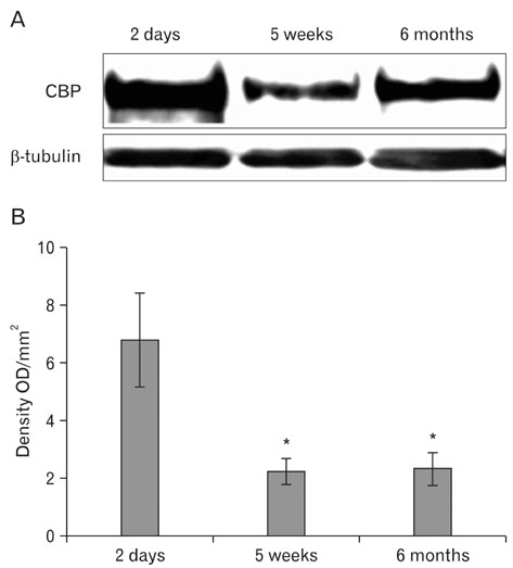
Fig. 1 Western blot analysis of cyclic AMP-responsive element binding protein-binding protein (CBP) in the retinas of 2-day-old, 5-week-old, and 6-month-old pigs. (A) A representative photograph of a Western blot for CBP and β-tubulin. CBP and β-tubulin are detected at adove 200 kDa and at approximately 55 kDa, respectively, as confirmed by molecular weight markers. (B) Bar graph of densitometric data analysis (mean±standard error, n=4-5 pigs/group). The relative levels of CBP are calculated after normalization to β-tubulin bands. The value indicating CBP expression levels from the retina at 2-day-old is arbitrarily defined as 100 (B, bar graphs). *P<0.05 vs. 2-day-old piglets.
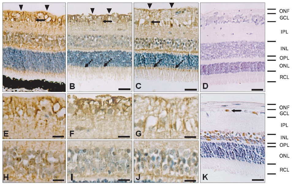
Fig. 2 Immunohistochemical analysis of cyclic AMP-responsive element binding protein-binding protein (CBP) in the retinas of 2-day-old (A, E, H), 5-week-old (B, F, L) and 6-month-old pigs (C, G, J). CBP immunoreactivity is detected in glial processes (arrowheads) and ganglion cells (arrow) in the ONF and GCL, some glial processes in the IPL, neuronal cells in INL, OPL, and RCL in the retinas of 2-day-old (A, E, H), 5-week-old (B, F, L) and 6-month-old pigs (C, G, J). (E and H, F and I, and G and J) are high-magnification images of GCL, and INL in (A, B, and C), respectively. No specific reaction product is seen in sections incubated with non-immune sera (D). In the rat retina, CBP immunostaining is evident in the ganglion cells (arrow) in the GCL, and some cells in the INL (K). Note the localization of CBP immunoreactivity mainly in the cytoplasm, the nucleus in the pig, and the retina in the rat. The retinal layers are indicated to the right: ONF, optic nerve fiber layer; GCL, ganglion cell layer; IPL, inner plexiform layer; INL, inner nuclear layer; OPL, outer plexiform layer; ONL, outer nuclear layer; RCL, layer of rods and cones. Counterstained with hematoxylin. Scale bars=25 µm (A-D, K), 12 µm (E-J).
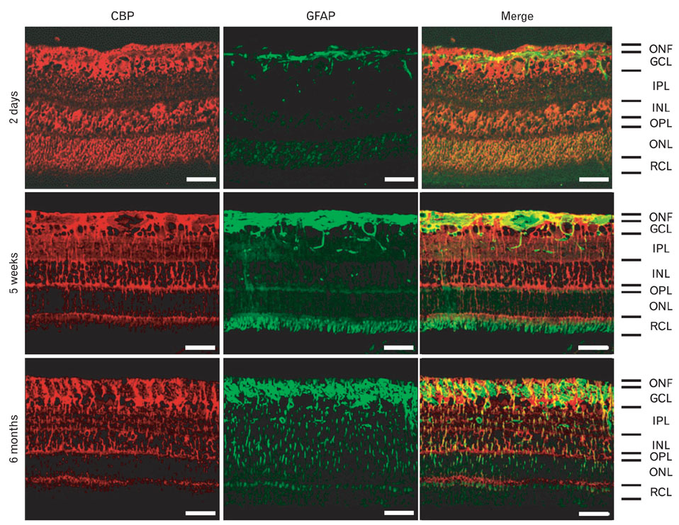
Fig. 3 Immunofluorescence staining of cyclic AMP-responsive element binding protein-binding protein (CBP, TRITC) with glial fibrillary acidic protein (GFAP, FITC) in the retina of 2-day-old, 5-week-old, and 6-month-old pigs. GFAP is detected very rarely in the retina of 2-day-old piglets, and it is abundant in the retina of 5-week-old and 6-month-old pigs. CBP immunoreactivity is occasionally co-localized in the GFAP-positive glial cells. ONF, optic nerve fiber layer; GCL, ganglion cell layer; IPL, inner plexiform layer; INL, inner nuclear layer; OPL, outer plexiform layer; ONL, outer nuclear layer; RCL, layer of rods and cones. Scale bars=50 µm.
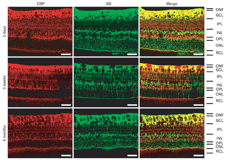
Fig. 4 Immunofluorescence staining of cyclic AMP-responsive element binding protein-binding protein (CBP, TRITC) with glutamine synthetase (GS, FITC) in the retina of 2-day-old, 5-week-old, and 6-month-old pigs. In ganglion cell layer (GCL), GS immunoreactivity is more abundant in retina of 2-day-old piglets compared to that of 5-week-old and 6-month-old pigs. CBP immunofluorescence is co-localized in GS-positive Müller cells. ONF, optic nerve fiber layer; IPL, inner plexiform layer; INL, inner nuclear layer; OPL, outer plexiform layer; ONL, outer nuclear layer; RCL, layer of rods and cones. Scale bars=50 µm.
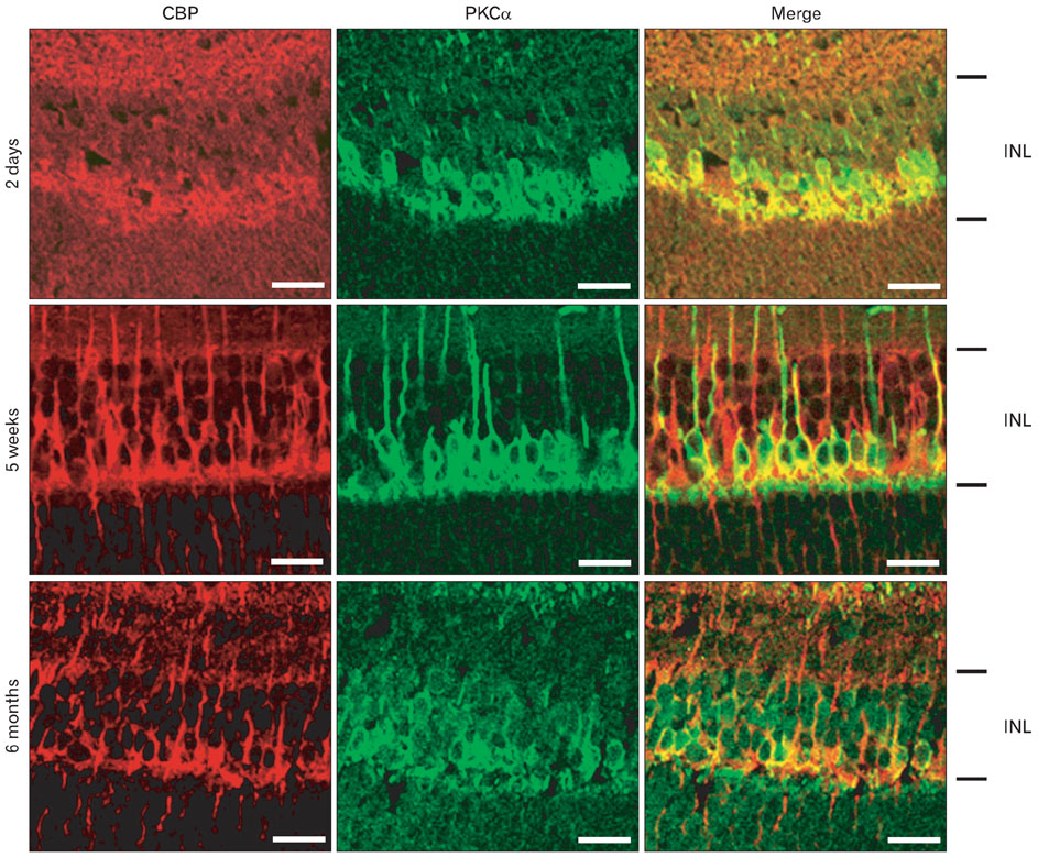
Fig. 5 Immunofluorescence staining of cyclic AMP-responsive element binding protein binding protein (CBP, TRITC) with protein kinase C, alpha (PKCα, FITC) in the inner nuclear layer (INL) of retina of 2-day-old, 5-week-old, and 6-month-old pigs. CBP immunofluorescence is co-localized in PKCα-positive bipolar cells. INL, inner nuclear layer. Scale bars=20 µm.
Reference
-
1. Brindle PK, Montminy MR. The CREB family of transcription activators. Curr Opin Genet Dev. 1992. 2:199–204.2. Chrivia JC, Kwok RP, Lamb N, Hagiwara M, Montminy MR, Goodman RH. Phosphorylated CREB binds specifically to the nuclear protein CBP. Nature. 1993. 365:855–859.3. Cohen HY, Lavu S, Bitterman KJ, Hekking B, Imahiyerobo TA, Miller C, Frye R, Ploegh H, Kessler BM, Sinclair DA. Acetylation of the C terminus of Ku70 by CBP and PCAF controls Bax-mediated apoptosis. Mol Cell. 2004. 13:627–638.4. Li Q, Xiao H, Isobe K. Histone acetyltransferase activities of cAMP-regulated enhancer-binding protein and p300 in tissues of fetal, young, and old mice. J Gerontol A Biol Sci Med Sci. 2002. 57:B93–B98.5. Wood MA, Kaplan MP, Park A, Blanchard EJ, Oliveira AM, Lombardi TL, Abel T. Transgenic mice expressing a truncated form of CREB-binding protein (CBP) exhibit deficits in hippocampal synaptic plasticity and memory storage. Learn Mem. 2005. 12:111–119.6. Rouaux C, Jokic N, Mbebi C, Boutillier S, Loeffler JP, Boutillier AL. Critical loss of CBP/p300 histone acetylase activity by caspase-6 during neurodegeneration. EMBO J. 2003. 22:6537–6549.7. Fiore P, Gannon RL. Expression of the transcriptional coactivators CBP and p300 in the hamster suprachiasmatic nucleus: possible molecular components of the mammalian circadian clock. Brain Res Mol Brain Res. 2003. 111:1–7.8. Jin K, Mao XO, Simon RP, Greenberg DA. Cyclic AMP response element binding protein (CREB) and CREB binding protein (CBP) in global cerebral ischemia. J Mol Neurosci. 2001. 16:49–56.9. Anderson J, Bhandari R, Kumar JP. A genetic screen identifies putative targets and binding partners of CREB-binding protein in the developing Drosophila eye. Genetics. 2005. 171:1655–1672.10. Ruiz-Ederra J, García M, Hernández M, Urcola H, Hernández-Barbáchano E, Araiz J, Vecino E. The pig eye as a novel model of glaucoma. Exp Eye Res. 2005. 81:561–569.11. Kim J, Moon C, Ahn M, Joo HG, Jin JK, Shin T. Immunohistochemical localization of galectin-3 in the pig retina during postnatal development. Mol Vis. 2009. 15:1971–1976.12. Lee J, Kim H, Lee JM, Shin T. Immunohistochemical localization of heat shock protein 27 in the retina of pigs. Neurosci Lett. 2006. 406:227–231.13. Kikuchi M, Tenneti L, Lipton SA. Role of p38 mitogen-activated protein kinase in axotomy-induced apoptosis of rat retinal ganglion cells. J Neurosci. 2000. 20:5037–5044.14. Harman A, Abrahams B, Moore S, Hoskins R. Neuronal density in the human retinal ganglion cell layer from 16-77 years. Anat Rec. 2000. 260:124–131.15. Dietze EC, Bowie ML, Mrózek K, Caldwell LE, Neal C, Marjoram RJ, Troch MM, Bean GR, Yokoyama KK, Ibarra CA, Seewaldt VL. CREB-binding protein regulates apoptosis and growth of HMECs grown in reconstituted ECM via laminin-5. J Cell Sci. 2005. 118(Pt 21):5005–5022.16. Johansson K, Törngren M, Wasselius J, Månsson L, Ehinger B. Developmental expression of DCC in the rat retina. Brain Res Dev Brain Res. 2001. 130:133–138.17. Bouchard JF, Moore SW, Tritsch NX, Roux PP, Shekarabi M, Barker PA, Kennedy TE. Protein kinase A activation promotes plasma membrane insertion of DCC from an intracellular pool: a novel mechanism regulating commissural axon extension. J Neurosci. 2004. 24:3040–3050.18. Moore SW, Kennedy TE. Protein kinase A regulates the sensitivity of spinal commissural axon turning to netrin-1 but does not switch between chemoattraction and chemorepulsion. J Neurosci. 2006. 26:2419–2423.19. Liu X, Green CB. Circadian regulation of nocturnin transcription by phosphorylated CREB in Xenopus retinal photoreceptor cells. Mol Cell Biol. 2002. 22:7501–7511.
- Full Text Links
-
- Actions
-
Cited
- CITED
-
- Close
- Share
-
- Similar articles
-
- Effects of dopamine and haloperidol on morphine-induced CREB and AP-1 DNA binding activities in differentiated SH-SY5Y human neuroblastoma cells
- β-Catenin Regulates the Expression of cAMP Response Element-Binding Protein 1 in Squamous Cell Carcinoma Cells
- A Functional Role for CREB as a Positive Regulator of Memory Formation and LTP
- Striatal CREB Phosphorylation Following Cued/Response Learning in the Water Maze Differ in Two Inbred Strains of Mice
- Developmental Changes in the Phosphorylation of CREB Following Electroconvulsive Shock in Rat Brain
