Method for Automatic Tube Current Selection for Obtaining a Consistent Image Quality and Dose Optimization in a Cardiac Multidetector CT
- Affiliations
-
- 1Department of Radiology, Peking University People's Hospital, China. cyanage8@gmail.com
- 2CT Imaging Research Center, GE Healthcare, China.
- KMID: 1102559
- DOI: http://doi.org/10.3348/kjr.2009.10.6.568
Abstract
OBJECTIVE
To evaluate a quantitative method for individually adjusting the tube current to obtain images with consistent noise in electrocardiogram (ECG)-gated CT cardiac scans. MATERIALS AND METHODS: The image noise from timing bolus and cardiac CT scans of 80 patients (Group A) who underwent a 64-row multidetector (MD) CT cardiac examination with patient-independent scan parameters were analyzed. A formula was established using the noise correlation between the timing bolus and cardiac scans. This formula was used to predict the required tube current to obtain the desired cardiac CT image noise based on the timing bolus noise measurement. Subsequently, 80 additional cardiac patients (Group B) were scanned with individually adjusted tube currents using an established formula to evaluate its ability to obtain accurate and consistent image noise across the patient population. Image quality was evaluated using score scale of 1 to 5 with a score of 3 or higher being clinically acceptable. RESULTS: Using the formula, we obtained an average CT image noise of 28.55 Hounsfield unit (HU), with a standard deviation of only 1.7 HU, as opposed to a target value of 28 HU. Image quality scores were 4.03 and 4.27 for images in Groups A and B, respectively, and there was no statistical difference between the image quality scores between the two groups. However, the average CT dose index (CTDIvol) was 30% lower for Group B. CONCLUSION: Adjusting the tube current based on timing bolus scans may provide a consistent image quality and dose optimization for cardiac patients of various body mass index values.
Keyword
MeSH Terms
Figure
-
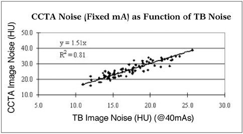
Fig. 1 Noise correlation between coronary CT angiography (CCTA) and timing bolus (TB) scans, both acquired at constant tube currents, showing good linear relationship.
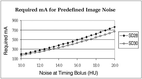
Fig. 2 Required tube current as function of timing bolus noise for same patient to obtain preset image noise levels of 28 HU and 30 HU.

Fig. 3 Scatter plot showing relationship between coronary CT angiography (CCTA) image noise and patient body mass index (BMI) for patients in Group A with constant mAs. Image noise in this group had wide distribution: 56 mAs to 245 mAs.
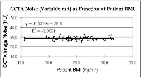
Fig. 4 Scatter plot showing relationship between coronary CT angiography image noise and patient body mass index (BMI) for patients in Group B with adaptive tube current setting. Image noise deviated very little from mean value of 28.6 HU.
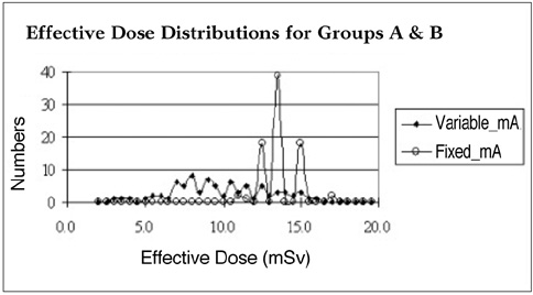
Fig. 5 Effective dose distributions for patients in groups A with constant mA and B, and with adaptive mA.
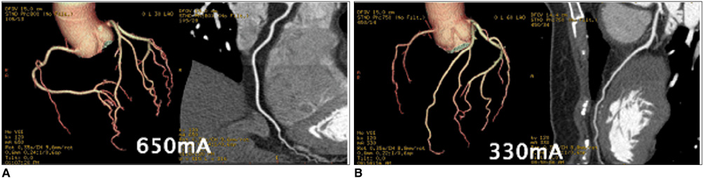
Fig. 6 Example of coronary CT angiography images with fixed (650 mA, left) and adaptive mA (330 mA, right) for patients from Groups A and B, respectively. Body mass index and image noise were 30.1 kg/m2 and 28.5 HU for Group A and 23.4 kg/m2 and 26.9 HU for Group B. Both images were rated excellent (score of 5). However, for patient in Group B, where tube current was adjusted based on timing bolus noise measurement, dose saving of about 50% was achieved compared to when fixed mA was used.
Reference
-
1. Hsieh J, Pan T, Acharya KC, Shen Y, Woodford M. Non-uniform phase coded image reconstruction for cardiac CT. Radiology. 1999. 213:401.2. Taguchi K, Anno H. High temporal resolution for multislice helical computed tomography. Med Phys. 2000. 27:861–872.3. Vembar M, Garcia MJ, Heuscher DJ, Haberl R, Matthews D, Böhme GE, et al. A dynamic approach to identifying desired physiological phases for cardiac imaging using multislice spiral CT. Med Phys. 2003. 30:1683–1693.4. Choi HS, Choi BW, Choe KO, Choi D, Yoo KJ, Kim MI, et al. Pitfalls, artifacts, and remedies in multi-detector row CT coronary angiography. Radiographics. 2004. 24:787–800.5. Bruder H, Stierstorfer K, McCollough C, Raupach R, Petersilka M, Grasruck M, et al. Design considerations in cardiac CT. Proc SPIE. 2006. 6142:61420.6. Haaga JR. Radiation dose management: weighing risk versus benefit. AJR Am J Roentgenol. 2001. 177:289–291.7. Slovis TL. CT and computed radiography: the pictures are great, but is the radiation dose greater than required. AJR Am J Roentgenol. 2002. 179:39–41.8. Hsieh J, Londt J, Vass M, Li J, Tang X, Okerlund D. Step-and-shoot data acquisition and reconstruction for cardiac x-ray computed tomography. Med Phys. 2006. 33:4236–4248.9. Hurwitz LM, Reiman RE, Yoshizumi TT, Goodman PC, Toncheva G, Nguyen G, et al. Radiation dose from contemporary cardiothoracic multidetector CT protocols with an anthropomorphic female phantom: implications for cancer induction. Radiology. 2007. 245:742–750.10. GB Avinash . Method and apparatus for enhancing discrete pixel images. US Patent. 6,208,763 B1. 2001.11. Jakobs TF, Becker CR, Ohnesorge B, Flohr T, Suess C, Schoepf UJ, et al. Multislice helical CT of the heart with retrospective ECG gating: reduction of radiation exposure by ECG-controlled tube current modulation. Eur Radiol. 2002. 12:1081–1086.12. Li J, Mohr K, Okerlund D. Dose reduction for CT coronary artery imaging using a special bowtie. Med Phys. 2004. 31:1841.13. McCollough CH, Bruesewitz MR, Kofler JM Jr. CT dose reduction and dose management tools: overview of available options. Radiographics. 2006. 26:503–512.14. Kalra MK, Maher MM, Toth TL, Schmidt B, Westerman BL, Morgan HT, et al. Techniques and applications of automatic tube current modulation for CT. Radiology. 2004. 233:649–657.15. Rizzo S, Kalra M, Schmidt B, Dalal T, Suess C, Flohr T, et al. Comparison of angular and combined automatic tube current modulation techniques with constant tube current CT of the abdomen and pelvis. AJR Am J Roentgenol. 2006. 186:673–679.16. Wilting JE, Zwartkruis A, van Leeuwen MS, Timmer J, Kamphuis AG, Feldberg M. A rational approach to dose reduction in CT: individualized scan protocols. Eur Radiol. 2001. 11:2627–2632.17. Mahnken AH, Wildberger JE, Simon J, Koos R, Flohr TG, Schaller S, et al. Detection of coronary calcifications: feasibility of dose reduction with a body weight-adapted examination protocol. AJR Am J Roentgenol. 2003. 181:533–538.18. Li J, Gao J, Sun X. How to obtain consistent cardiac CT image noise for patients of different sizes: body mass index based tube current selection. RSNA Abstract Book. 2007. 730.19. General principles associated with good imaging technique: technical, clinical and physical parameters. Accessed in August 28, 2009. http://www.drs.dk/guidelines/ct/quality/mainindex.htm. Chapter 1, page 4.20. Das M, Mahnken AH, Mühlenbruch G, Stargardt A, Weiss C, Sennst DA, et al. Individually adapted examination protocols for reduction of radiation exposure for 16-MDCT chest examinations. AJR Am J Roentgenol. 2005. 184:1437–1443.21. Hur G, Hong SW, Kim SY, Kim YH, Hwang YJ, Lee WR, et al. Uniform image quality achieved by tube current modulation using SD of attenuation in coronary CT angiography. AJR Am J Roentgenol. 2007. 189:188–196.22. Fei X, Du X, Li P, Liao J, Shen Y, Li K. Effect of dose-reduced scan protocols on cardiac coronary image quality with 64-row MDCT: a cardiac phantom study. Eur J Radiol. 2008. 67:85–91.
- Full Text Links
-
- Actions
-
Cited
- CITED
-
- Close
- Share
-
- Similar articles
-
- Is It Better to Enter a Volume CT Dose Index Value before or after Scan Range Adjustment for Radiation Dose Optimization of Pediatric Cardiothoracic CT with Tube Current Modulation?
- Combined Use of Automatic Tube Voltage Selection and Current Modulation with Iterative Reconstruction for CT Evaluation of Small Hypervascular Hepatocellular Carcinomas: Effect on Lesion Conspicuity and Image Quality
- Review of the Asian Consortium on Radiation Dose of Pediatric Cardiac CT (ASCI-REDCARD) and Recommendations for a New Edition
- Effects of Iterative Reconstruction Algorithm, Automatic Exposure Control on Image Quality, and Radiation Dose: Phantom Experiments with Coronary CT Angiography Protocols
- Attenuation-Based Automatic Tube Potential Selection in Cerebral Computed Tomography Angiography: Effects on Radiation Exposure and Image Quality
