Attenuation-Based Automatic Tube Potential Selection in Cerebral Computed Tomography Angiography: Effects on Radiation Exposure and Image Quality
- Affiliations
-
- 1Department of Radiology, Gangnam Severance Hospital, Yonsei University College of Medicine, Seoul, Korea. jjondol@yuhs.ac
- KMID: 2399304
- DOI: http://doi.org/10.3348/jksr.2018.78.1.35
Abstract
OBJECTIVE
To investigate the feasibility of using the attenuation-based automatic tube potential selection (ATPS) algorithm for cerebral computed tomography angiography (CTA) and to assess radiation dose, vascular attenuation, and image quality compared to a conventional fixed 120-kVp protocol.
MATERIALS AND METHODS
Among 36 volunteers for cerebral CTA, a total of 18 were scanned with fixed 120 kVp and 140 effective mAs using automatic tube current modulation. The other 18 were scanned with an ATPS algorithm. Radiation doses, attenuation, contrast-to-noise ratio (CNR) of the cerebral arteries, subjective scores for arterial attenuation, edge sharpness of the artery, visibility of small arteries, venous contamination, image noise, and overall image quality were compared between the groups.
RESULTS
The volume CT dose index and effective dose of the ATPS group were lower than those of the fixed 120-kVp group. The ATPS group had significantly higher arterial attenuation and no significant difference in CNR, compared with the fixed 120-kVp. The ATPS group had higher subjective scores for arterial attenuation, edge sharpness of the artery, visibility of small arteries, and overall image quality.
CONCLUSION
The ATPS algorithm for the cerebral CTA reduced radiation dose by 43% while maintaining image quality and improved the attenuation of cerebral arteries by selecting lower tube potential.
MeSH Terms
Figure
-

Fig. 1. Cerebral CTA of 38-year-old woman, using the fixed 120-kVp protocol. Axial (A) and coronal (B) maximum intensity projection images with a slab thickness of 10 mm and volume rendering image (C) were assessed. The mean attenuation value of the cerebral arteries (397.5 HU) and CNR (25.5) in this volunteer were similar to the mean attenuation value (377.3 HU) and mean CNR (24.7) for cerebral CTA in group A. CNR = contrast-to-noise radio, CTA = computed tomography angiography, HU = Hounsfield units
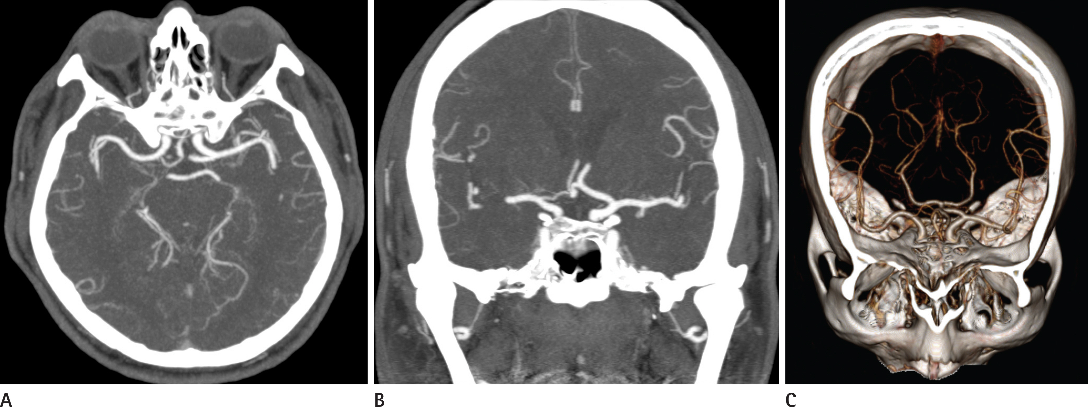
Fig. 2. Cerebral CTA of 50-year-old man, using the automatic tube potential selection protocol. Axial (A) and coronal (B) maximum intensity projection images with a slab thickness of 10 mm and volume rendering image (C) were assessed. A tube potential of 80 kVp was selected. The mean attenuation value of the cerebral arteries (560.5 HU) and CNR (22.8) in this volunteer were similar to the mean attenuation value (587.7 HU) and mean CNR (24.2) for cerebral CTA in group B. CNR = contrast-to-noise radio, CTA = computed tomography angiography, HU = Hounsfield units
Reference
-
References
1. Lell MM, Anders K, Uder M, Klotz E, Ditt H, Vega-Higuera F, et al. New techniques in CT angiography. Radiographics. 2006; 26(Suppl 1):S45–S62.
Article2. Berrington de González A, Mahesh M, Kim KP, Bhargavan M, Lewis R, Mettler F, et al. Projected cancer risks from computed tomographic scans performed in the United States in 2007. Arch Intern Med. 2009; 169:2071–2077.
Article3. Brenner DJ, Hall EJ. Computed tomography–an increasing source of radiation exposure. N Engl J Med. 2007; 357:2277–2284.4. Rizzo S, Kalra M, Schmidt B, Dalal T, Suess C, Flohr T, et al. Comparison of angular and combined automatic tube current modulation techniques with constant tube current CT of the abdomen and pelvis. AJR Am J Roentgenol. 2006; 186:673–679.
Article5. Mulkens TH, Bellinck P, Baeyaert M, Ghysen D, Van Dijck X, Mussen E, et al. Use of an automatic exposure control mechanism for dose optimization in multi-detector row CT examinations: clinical evaluation. Radiology. 2005; 237:213–223.
Article6. McCollough CH, Bruesewitz MR, Kofler JM Jr. CT dose reduction and dose management tools: overview of available options. Radiographics. 2006; 26:503–512.
Article7. Gunn ML, Kohr JR. State of the art: technologies for computed tomography dose reduction. Emerg Radiol. 2010; 17:209–218.
Article8. Cho ES, Chung TS, Oh DK, Choi HS, Suh SH, Lee HK, et al. Cerebral computed tomography angiography using a low tube voltage (80 kVp) and a moderate concentration of iodine contrast material: a quantitative and qualitative comparison with conventional computed tomography angiography. Invest Radiol. 2012; 47:142–147.9. Waaijer A, Prokop M, Velthuis BK, Bakker CJ, de Kort GA, van Leeuwen MS. Circle of Willis at CT angiography: dose reduction and image quality–reducing tube voltage and increasing tube current settings. Radiology. 2007; 242:832–839.
Article10. Bahner ML, Bengel A, Brix G, Zuna I, Kauczor HU, Delorme S. Improved vascular opacification in cerebral computed tomography angiography with 80 kVp. Invest Radiol. 2005; 40:229–234.
Article11. Szucs-Farkas Z, Schaller C, Bensler S, Patak MA, Vock P, Schindera ST. Detection of pulmonary emboli with CT angiography at reduced radiation exposure and contrast material volume: comparison of 80 kVp and 120 kVp protocols in a matched cohort. Invest Radiol. 2009; 44:793–799.12. Schindera ST, Graca P, Patak MA, Abderhalden S, von All-men G, Vock P, et al. Thoracoabdominal-aortoiliac multidetector-row CT angiography at 80 and 100 kVp: assessment of image quality and radiation dose. Invest Radiol. 2009; 44:650–655.13. Heyer CM, Mohr PS, Lemburg SP, Peters SA, Nicolas V. Image quality and radiation exposure at pulmonary CT angiography with 100- or 120-kVp protocol: prospective randomized study. Radiology. 2007; 245:577–583.
Article14. Schindera ST, Nelson RC, Mukundan S Jr, Paulson EK, Jaffe TA, Miller CM, et al. Hypervascular liver tumors: low tube voltage, high tube current multi-detector row CT for enhanced detection–phantom study. Radiology. 2008; 246:125–132.
Article15. Kalra MK, Maher MM, Toth TL, Hamberg LM, Blake MA, Shepard JA, et al. Strategies for CT radiation dose optimization. Radiology. 2004; 230:619–628.
Article16. Brix G, Lechel U, Petersheim M, Krissak R, Fink C. Dynamic contrast-enhanced CT studies: balancing patient exposure and image noise. Invest Radiol. 2011; 46:64–70.17. Chen GZ, Zhang LJ, Schoepf UJ, Wichmann JL, Milliken CM, Zhou CS, et al. Radiation dose and image quality of 70 kVp cerebral CT angiography with optimized sinogram-affirmed iterative reconstruction: comparison with 120 kVp cerebral CT angiography. Eur Radiol. 2015; 25:1453–1463.
Article18. Komlosi P, Zhang Y, Leiva-Salinas C, Ornan D, Patrie JT, Xin W, et al. Adaptive statistical iterative reconstruction reduces patient radiation dose in neuroradiology CT studies. Neuroradiology. 2014; 56:187–193.
Article19. Löve A, Siemund R, Höglund P, Ramgren B, Undrén P, Björk-man-Burtscher IM. Hybrid iterative reconstruction algorithm improves image quality in craniocervical CT angiography. AJR Am J Roentgenol. 2013; 201:W861–W866.
Article20. Yu L, Li H, Fletcher JG, McCollough CH. Automatic selection of tube potential for radiation dose reduction in CT: a general strategy. Med Phys. 2010; 37:234–243.
Article21. Winklehner A, Goetti R, Baumueller S, Karlo C, Schmidt B, Raupach R, et al. Automated attenuation-based tube potential selection for thoracoabdominal computed tomography angiography: improved dose effectiveness. Invest Radiol. 2011; 46:767–773.22. Lee KH, Lee JM, Moon SK, Baek JH, Park JH, Flohr TG, et al. Attenuation-based automatic tube voltage selection and tube current modulation for dose reduction at contrast-enhanced liver CT. Radiology. 2012; 265:437–447.
Article23. Eller A, May MS, Scharf M, Schmid A, Kuefner M, Uder M, et al. Attenuation-based automatic kilovolt selection in abdominal computed tomography: effects on radiation exposure and image quality. Invest Radiol. 2012; 47:559–565.24. Ghoshhajra BB, Engel LC, Károlyi M, Sidhu MS, Wai B, Barreto M, et al. Cardiac computed tomography angiography with automatic tube potential selection: effects on radiation dose and image quality. J Thorac Imaging. 2013; 28:40–48.25. Park YJ, Kim YJ, Lee JW, Kim HY, Hong YJ, Lee HJ, et al. Automatic Tube Potential Selection with Tube Current Modulation (APSCM) in coronary CT angiography: comparison of image quality and radiation dose with conventional body mass index-based protocol. J Cardiovasc Comput Tomogr. 2012; 6:184–190.
Article26. Krazinski AW, Meinel FG, Schoepf UJ, Silverman JR, Canstein C, De Cecco CN, et al. Reduced radiation dose and improved image quality at cardiovascular CT angiography by automated attenuation-based tube voltage selection: intra-individual comparison. Eur Radiol. 2014; 24:2677–2684.
Article27. Spearman JV, Schoepf UJ, Rottenkolber M, Driesser I, Canstein C, Thierfelder KM, et al. Effect of automated attenuation-based tube voltage selection on radiation dose at CT: an observational study on a global scale. Radiology. 2016; 279:167–174.
Article28. Furtado AD, Adraktas DD, Brasic N, Cheng SC, Ordovas K, Smith WS, et al. The triple rule-out for acute ischemic stroke: imaging the brain, carotid arteries, aorta, and heart. AJNR Am J Neuroradiol. 2010; 31:1290–1296.
Article29. Ertl-Wagner BB, Hoffmann RT, Bruning R, Herrmann K, Snyder B, Blume JD, et al. Multi-detector row CT angiography of the brain at various kilovoltage settings. Radiology. 2004; 231:528–535.
Article30. Commission of the European Community. European guidelines on quality criteria for computed tomography. Report EUR 16262 EN, 1999. Available at:. http://www.drs.dk/guidelines/ct/quality/htmlindex.htm. Accessed May 28,. 2017.31. Landis JR, Koch GG. The measurement of observer agreement for categorical data. Biometrics. 1977; 33:159–174.
Article32. Cho ES, Chung TS, Ahn SJ, Chong K, Baek JH, Suh SH. Cerebral computed tomography angiography using a 70 kVp protocol: improved vascular enhancement with a reduced volume of contrast medium and radiation dose. Eur Radiol. 2015; 25:1421–1430.
Article33. Curry TS, Murry RC. Basic interactions between X-rays and matter. In Curry TS, Dowdey JE, Murry RC, eds. Christensen's physics of diagnostic radiology. 4th ed.Philadelphia, PA: Lippincott Williams & Wilkins;2006. p. 61–69.34. Niemann T, Henry S, Faivre JB, Yasunaga K, Bendaoud S, Simeone A, et al. Clinical evaluation of automatic tube voltage selection in chest CT angiography. Eur Radiol. 2013; 23:2643–2651.
Article35. Mayer C, Meyer M, Fink C, Schmidt B, Sedlmair M, Schoenberg SO, et al. Potential for radiation dose savings in abdominal and chest CT using automatic tube voltage selection in combination with automatic tube current modulation. AJR Am J Roentgenol. 2014; 203:292–299.
Article36. Bushby KM, Cole T, Matthews JN, Goodship JA. Centiles for adult head circumference. Arch Dis Child. 1992; 67:1286–1287.
Article
- Full Text Links
-
- Actions
-
Cited
- CITED
-
- Close
- Share
-
- Similar articles
-
- Attenuation-Based Automatic Kilovoltage Selection and Sinogram-Affirmed Iterative Reconstruction: Effects on Radiation Exposure and Image Quality of Portal-Phase Liver CT
- Effects of Iterative Reconstruction Algorithm, Automatic Exposure Control on Image Quality, and Radiation Dose: Phantom Experiments with Coronary CT Angiography Protocols
- Combined Use of Automatic Tube Voltage Selection and Current Modulation with Iterative Reconstruction for CT Evaluation of Small Hypervascular Hepatocellular Carcinomas: Effect on Lesion Conspicuity and Image Quality
- Radiation Dose Reduction without Compromise to Image Quality by Alterations of Filtration and Focal Spot Size in Cerebral Angiography
- Method for Automatic Tube Current Selection for Obtaining a Consistent Image Quality and Dose Optimization in a Cardiac Multidetector CT
